Triple Trouble: Colonic Obstruction by Sigmoid Volvulus in a Patient Affected by Ogilvie Syndrome and Giant Bochdalek Hernia
A B S T R A C T
Bochdalek hernia is a congenital left diaphragmatic hernia (CDH), usually diagnosed in children following the onset of thoracic symptoms such as dyspnea or tachycardia. Ogilvie’s syndrome (or acute colonic pseudo-obstruction) may be due to several conditions, including neurologic diseases, neuroleptic therapy and systemic syndromes, and is characterized by massive colon dilation in the absence of mechanical obstruction or toxic mega-colon. Sigmoid volvulus, consisting of sigmoid rotation around its axis, is a relatively frequent cause of large bowel obstruction. The three simultaneous conditions in the same patient in an emergency setting are rare, leading to a challenging situation concerning diagnosis and management. Here, we report the case of a mentally disabled 59-year-old female presenting with colonic obstruction caused by sigmoid volvulus after several episodes of Ogilvie’s syndrome-related pseudo-occlusion, found to have a giant left-sided Bochdalek diaphragmatic hernia. The patient was treated by an emergency laparoscopic approach. Despite the resolution of the abdominal picture, the patient died on postoperative day 15 by respiratory failure.
Keywords
Colonic obstruction, sigmoid volvulus, Ogilvie syndrome, Bochdalek hernia
Introduction
Bochdalek hernia (BH) is a congenital diaphragmatic hernia caused by the incomplete fusion of the posterolateral diaphragmatic foramina during the 9-10th week of pregnancy, resulting in abdominal organs displacement into the thorax [1, 2]. Accounting for 0.17-6% of all diaphragmatic hernias, BH usually occurs on the left side 80-90%, is usually diagnosed at a young age, and may result in incarcerated bowel or severe pulmonary disease [3, 4]. Small or large bowel migration through the diaphragm into thorax is the most frequent cause of intestinal obstruction in BH patients [4]. Ogilvie syndrome (OS) consists of an acute pseudo-obstruction caused by colon dilatation without cause and is mostly reported in patients affected by neurologic or psychiatric disorders, under antipsychotic drugs such as clozapine causing gastrointestinal hypomobility [5]. Typically, bowel dilation is limited to proximal colon, being maximal at the cecum with a transition-zone near the splenic flexure [5, 6].
Colonic volvulus consists of the rotation of the colon around its mesentery, causing intestinal obstruction. It is the third most frequent cause of large bowel obstruction after cancer and diverticulitis in the developed world, accounting for less than 5% of large bowel obstructions [7, 8]. Sigmoid is frequently affected (60-75%), followed by cecum (25-40%), transverse colon (1-4%), and splenic flexure (1%) [9]. Sigmoid volvulus’s typical presentation includes abdominal distension, pain, and vomiting, whereas management is nowadays endoscopic derotation, with salvage surgery after unsuccessful endoscopy or ongoing colonic necrosis/perforation [10].
The association of colonic volvulus with various conditions, including pregnancy, chronic constipation, mobile cecum, and congenital redundant colon, has been described, while the association with mental retardation is rarer [11, 12]. Here we report the first case of colonic obstruction in a mentally disabled patient with OS and Bochdalek hernia. The peculiar issues of this case are discussed – how to achieve such a diagnosis in a non-collaborative patient, and, what is the adequate management of those concomitant conditions in an emergency setting.
Case Presentation
A 59-year-old female patient was admitted to the emergency room with colicky pain, dysphagia, constipation, and dyspnea. She was a long-time psychiatric hospital inpatient with severe schizophrenia and dementia on clozapine therapy already undergoing. multiple episodes of pseudo-occlusion managed conservatively, while no thoracic/abdominal trauma was reported. Laboratory tests showed leukocytosis (WBC 16 x103/µL), elevated protein chain reaction (PCR) serum level (264 mg/L), and hypokalaemia. Arterial blood gas analysis showed hypercapnia and hypoxia. Physical examination elicited diffuse abdominal pain and abdominal distention with dyspnea. X-rays showed diffuse colonic dilatation, both in the abdomen and the thorax, reaching the projection line of the left collarbone. CT-scan (Figure 1) showed a massive herniation of intra-abdominal organs into the left hemithorax through a large defect in the left posterior diaphragm, associated with diffuse colonic distension.
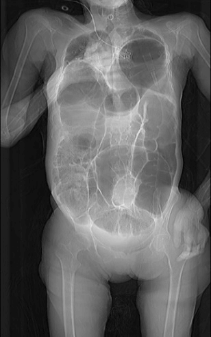
Figure 1: Computerized tomographic images showing distention of the entire colon.
After unsuccessful endoscopic derotation, emergency surgical exploration was performed by laparoscopy. Diaphragmatic hernia content was reduced in the abdomen, the sac was resected, and the prosthesis placed. At derotation, the sigmoid colon appeared hugely dilated and partly necrotic; therefore, the affected segment was resected, and terminal sigmoid ostomy, in a Hartmann procedure fashion, was performed. The postoperative course was uneventful with bowel movement on POD 4, until POD 15, when the patient developed dyspnea, rapidly deteriorating to respiratory failure until death occurred 6 hours later.
Discussion
This case raises two major questions – how to achieve the diagnosis of intestinal obstruction in a mentally impaired patient and which is the most appropriate management of those three concomitant conditions, namely sigmoid volvulus complicated by colonic segmental necrosis, Ogilvie syndrome and massive Bochdalek hernia in an emergency setting.
Diagnosis of acute abdomen in a mentally impaired patient is often challenging due to both non-specific clinical presentation and the obvious difficulty in the collection of symptoms and medical history. OS has been reported to be a long-term complication of psychotropic drug use, and Clozapine-induced gastrointestinal hypomotility (CRGH) is one supposed physio pathologic mechanism responsible not only of recurrent pseudo-obstruction episodes, but also chronic constipation [5]. Chronic constipation can cause lengthening of the colon, eventually leading to a redundant sigmoid colon, which is a prerequisite of sigmoid volvulus [13].
Typical sigmoid volvulus presentation is distal large bowel obstruction needing emergency management, but, rarely, its clinical presentation may be as a chronic recurrent abdominal pain [14]. In this light, it is questionable that the reported previous recurrent episodes of the then-called “pseudo-obstruction” in an OS context were not recurrent episodes of “real obstruction” caused by sigmoid volvulus. Sigmoid volvulus most common symptoms are abdominal pain (66%) and vomiting (31%), while more frequent signs are abdominal distention (69%) and tenderness (41%) [15, 16]. The diagnostic delay may lead to bowel ischemia, perforation, shock and multiorgan dysfunction leading to death. In the present case, at the time of surgical exploration, colonic necrosis was already present, and colonic resection was the only viable option.
Sigmoid volvulus may be difficult to diagnose in mentally disabled patients, where bowel distention may be the result of long-term aerophagia and constipation, thus increasing the difficulty of interpretation of clinical signs/symptoms as well as imaging findings. Radiological examinations play a pivotal role in the pre-operative diagnosis of such patients with impaired communication. Plain erect abdominal X-rays generally show a dilated sigmoid colon and a coffee bean-like shape formed by grossly dilated and closely apposed sigmoid loops [15, 16]. In the absence of an adequate physical examination, the only examination possibly suggesting that obstruction is evolving in colonic ischemia/necrosis is dosing the serum inflammatory markers level, including white blood cells and PCR.
In the present case, CT-scan and inflammatory markers rapidly assessed the nature and severity of obstruction, thus allowing for the prompt transfer to the operating theatre. Although the management of this episode is seemingly timely, it is debatable whether previous episodes of supposed pseudo-obstruction, where the patient did not undergo any laboratory nor imaging examination, has been correctly managed. Seemingly, in such a context of multiple conditions in a mentally impaired patient under medications, before labelling any new episode as recurrent pseudo-obstruction, blood test and CT-scan should be routinely performed to rule out any condition suitable of emergency or delayed surgery.
The main aim of treatment of sigmoid volvulus is to resolve bowel obstruction, thus avoiding ischemia/necrosis, and to prevent recurrence [17]. In non-necrotic non-perforated sigmoid volvulus, water-soluble contrast enema studies can be used for diagnostic and therapeutic purpose [15, 17]. In our experience, in hemodynamically stable patients with no feature of perforation, we associate barium enema with proctoscopy and sigmoid decompression by rectally introduced tube. The most common non-surgical method of sigmoid volvulus nowadays remains endoscopic decompression, which may be performed in most environments with a low morbidity rate [12]. Since a recurrence rate of 35-90% associated with the non-operative approach has been reported, delayed, non-emergency surgical management is considered by most surgeons [16-18]. Whenever colonic necrosis may not be excluded by examinations or non-operative maneuvers, surgical exploration is deemed necessary by open and laparoscopic approach. Depending on intraoperative findings, various surgical options, including derotation alone, derotation with colopexy, resection with primary anastomosis and resection with end-colostomy.
In the present case, we faced an even more complex scenario, with two additional challenging conditions: massive colonic distension, at least partly not caused by occlusion but by disease (or therapy)-induced colonic hypomobility, and a huge bowel herniation into the thorax through a large diaphragmatic hernia, probably worsening intestinal obstruction’s picture. Scholastically, the treatment of major diaphragmatic defects is prosthetic repair, which we eventually performed by laparoscopy without particular issues [19].
Although the patient intraoperatively well tolerated the procedure, it may be supposed that, after resolving the obstruction status, the presence of major distension of the colonic remnant may have played a role in postoperative respiratory breakdown and death. Probably, the mini-invasive approach (laparoscopic access, minor resection) has allowed an early improvement with bowel movement on POD 4. Possibly, during the following days, the pre-existing OS-related distension, worsened by herniated bowel complete reduction into the abdomen, altered respiratory dynamics by reducing diaphragmatic excursion during inspiratory-expiratory acts. Although it is impossible to draw any conclusive teaching from the present case, perhaps, a larger or subtotal colectomy may have allowed reducing intra-abdominal pressure, thus improving respiration, in the early postoperative course.
Conclusion
The occurrence of sigmoid volvulus in a mentally disabled patient as well as in a patient with concomitant OS and Bochdalek hernia may result in a challenging situation for the physician and the surgeon, concerning both accurate and timely diagnosis and adequate surgical treatment. In such a complex and misleading scenario, any new episode of possible intestinal obstruction should be carefully evaluated and adequately studied by blood sample and CT-scan. At surgical exploration, if a colonic resection is eventually indicated owing to ongoing segmental necrosis, a more extended colonic resection, or even a subtotal colectomy, may be a viable option in order to reduce the risk of postoperative respiratory failure.
Conflicts of Interest
None.
Funding
None.
Article Info
Article Type
Case ReportPublication history
Received: Mon 27, Apr 2020Accepted: Mon 25, May 2020
Published: Fri 05, Jun 2020
Copyright
© 2023 Renato Costi. This is an open-access article distributed under the terms of the Creative Commons Attribution License, which permits unrestricted use, distribution, and reproduction in any medium, provided the original author and source are credited. Hosting by Science Repository.DOI: 10.31487/j.SCR.2020.05.12
Figures & Tables
References
- Luther A, Mahajan A (2015) Left-sided Bochdalek Hernia in an Adult: A Case Report with Review of Literature. J Int Med Sci Acad 28: 33-34.
- Alam A, Chander BN (2005) Adult Bochdalek Hernia. Med J Armed Forces India 61: 284-286. [Crossref]
- Mullins ME, Stein J, Saini SS, Mueller PR (2001) Prevalence of Incidental Bochdalek's Hernia in a Large Adult Population. Am J Roentgenol 177: 363-366. [Crossref]
- Shin MS, Mulligan SA, Baxley WA Ho KJ (1987) Bochdalek Hernia of Diaphragm in the Adult. Diagnosis by Computed Tomography. Chest 92: 1098-1101. [Crossref]
- Akkaş M, Demir MC (2020) Clozapine Related Ogilvie Syndrome With Fatal Outcome. Riv Psichiatr 55: 53-56. [Crossref]
- Shahait AD, Mostafa G (2018) Ogilvie's Syndrome or Colonic Pseudo-Obstruction. Am Surg 84: e38-e39. [Crossref]
- Ballantyne GH, Brandner MD, Beart RW, Ilstrup DM (1985) Volvulus of the Colon. Incidence and Mortality. Ann Surg 202: 83-92. [Crossref]
- Bagarani M, Conde AS, Longo R, Italiano A, Terenzi A et al. (1993) Sigmoid Volvulus in West Africa: A Prospective Study on Surgical Treatments. Dis Colon Rectum 36: 186-190. [Crossref]
- Halabi WJ, Jafari MD, Kang CY, Nguyen VQ, Carmichael JC et al. (2014) Colonic Volvulus in the United States: Trends, Outcomes, and Predictors of Mortality. Ann Surg 259: 293-301. [Crossref]
- Gingold D, Murrell Z (2012) Management of Colonic Volvulus. Clin Colon Rectal Surg 25: 236- 244. [Crossref]
- Clermidi P, Abadie V, Campeotto F, Irtan S (2015) Sigmoid Volvulus: An Underestimated Cause of Intestinal Obstruction in Cornelia De Lange Syndrome. J Pediatr 167: 941. [Crossref]
- Frischman WJ, Couper RT, Freeman JK (1996) Cecal Volvulus Following Gastroduodenoscopy in Cornelia De Lange Syndrome. J Pediatr Gastroenterol Nutr 22: 205-207. [Crossref]
- Ismail A (1997) Recurrent Colonic Volvulus in Children. J Pediatr Surg 32: 1739-1742. [Crossref]
- Samuel M, Boddy SA, Nicholls E, Capps S (2000) Large Bowel Volvulus in Childhood. Aust N Z J Surg 70: 258-262. [Crossref]
- Lin MP, Chen YL, Tzeng WS (2011) Diagnosis of Sigmoid Volvulus Using the Coffee Bean, Northern Exposure Sign, Whirl Sign and Transition Point. BMJ Case Rep 2011: 0620114334. [Crossref]
- Perrot L, Fohlen A, Alves AJ, Lubrano J (2016) Management of the Colonic Volvulus in 2016. J Visc Surg 153: 183-192. [Crossref]
- Parolini F, Orizio P, Bulotta AL, Magne MG, Boroni G et al. (2016) Endoscopic Management of Sigmoid Volvulus in Children. World J Gastrointest Endosc 8: 439-443. [Crossref]
- Colinet S, Rebeuh J, Gottrand F, Kalach N, Paquot I et al. (2015) Presentation and Endoscopic Management of Sigmoid Volvulus in Children. Eur J Pediatr 174: 965-969. [Crossref]
- Puligandla P, Skarsgard E, Offringa M, Adatia I, Baird R et al. (2018) Diagnosis and Management of Congenital Diaphragmatic Hernia: A Clinical Practice Guideline. CMAJ 190: E103-E112. [Crossref]