
Transferring Mesenchymal Stem Cells to the Osteoarthritic Cartilage: Translating Science to Medicine
A B S T R A C T
Osteoarthritis (OA) is associated with loss of the phenotypic stability of affected joints. Due to its avascular nature, biomechanical properties of the cartilage tissue in a synovial joint are particularly compromised as the disease progresses. Current therapies for OA merely buy time until arthrodesis or arthroplasty, the ‘end game’, is ultimately reached. Over the last two decades, promising experimental data has started to emerge on mesenchymal stem cells (MSCs) as candidate cell types for cell and/ or tissue-based cartilage tissue repair. The principal attraction of MSCs lies in their consistent chondrogenic differentiation and proliferative propensities. Practically, they offer ease of isolation and culture-expansion. Targeted gene therapy can further enhance their delivery into osteoarthritic joints. Henceforth, understanding the biological activities and mechanisms of action of MSCs is crucial for a rational approach to their clinical application in osteoarthritic joints.
Keywords
Mesenchymal stem cells, osteoarthritis, cartilage, cell-based therapy, immuno-modulation
Basic Science of Articular Cartilage
The articular cartilage is a whitish, glassy substance that lines synovial joints. It is composed of specialised cells called chondrocytes, embedded in an extracellular matrix of collagen fibers, proteoglycan, and elastin fibers [1]. The interaction between the negatively charged cartilage proteoglycans notably, aggrecan and type II collagen provides the compressive and tensile strength of the joint tissue. Mechanical injury to the articular cartilage secondary to osteoarthritis (OA) causes significant musculoskeletal morbidity.
Articular Cartilage and Osteoarthritis
The basic pathophysiological feature of OA is the loss of compliance and strength of articular cartilage to allow for its function. Although they make up only 2% of the weight of articular cartilage, chondrocytes are the key elements in the development of the osteoarthritic process, due to their relatively inert nature and little regenerative capacity [2, 3]. As such, recent studies into mechanisms underpinning articular cartilage damage in OA have primarily focused on chondrocyte apoptosis [4, 5].
A study by Sharif et al. concluded that the degree of chondrocyte apoptosis was in fact significantly correlated with articular cartilage fibrillation and cellularity [5]. Gupta et al. postulated that as OA progresses, intraarticular chondrocytes synthesize and secrete proteolytic matrix metalloproteinases (MMPs) and aggrecanases [6]. These further recruit and activate the main pro-inflammatory cytokines, tumor necrosis factor (TNF)-a and interleukin (IL)-1. These two cytokines exhibit synergism; working together to amplify the process of articular cartilage degradation and apoptotic chondrocyte death.
Current Therapies for Cartilage Restoration in Osteoarthritis
Current procedures for OA cartilage restoration include autologous chondrocyte implantation, micro-fracture (Figure 1) and osteochondral transplantation (using allografts and autografts) [7-9]. In general, their goal is to stimulate ‘new’ articular cartilage growth. However, long, open, and deep incisions are often required, exposing patients to surgical or procedure- site infections.
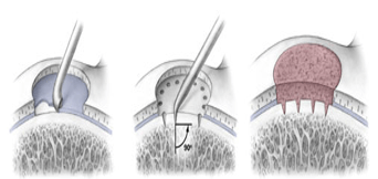
Figure 1: Shows the steps of the micro-fracture technique (schematic diagram). A) Damaged cartilage is removed, B) An awl is used to make holes in the subchondral bone, C) Healing response brings regenerative and healthy cartilage cells [10].
Secondly and more importantly, these techniques are limited to repair of focal cartilage lesions and as such, patients with extensive articular injury as seen in moderate and severe OA, are often excluded from such current treatment regimes, as they tend not to benefit much. Essentially, the inadequacies with current cartilage-repair therapies means that, perhaps, investing in MSCs-based research may be the most effective approach of arresting OA’s pathogenesis both in the early and advanced stages of the disease process.
Biology of Mesenchymal Stem Cells
The International Society for Cellular Therapy (ISCT) proposes three criteria to define mesenchymal stem cells (MSCs) and their characteristics:
i. Adherence to plastic.
ii. Specific surface antigen (Ag) expression.
iii.Multipotent differentiation potential [11].
Originally isolated from bone marrow by Frendeistein et al. MSCs can be easily isolated from the umbilical cord, cartilage, muscle, and tendon [12]. They are then engineered and ‘expanded’ in a culture media and used to produce a diverse range of cartilaginous skeletal tissues.
Immuno-Modulatory Properties of MSCs
A key feature of MSCs is their hypo/ non-immunogenic nature. Greene et al. explained that, as OA progresses, the pro-inflammatory cytokines recruit more pro-inflammatory mediators as well as MSCs (‘anti-inflammatory cytokines’) to areas of the inflammation within the cartilage defect [13].
Activated MSCs, once within the cartilage, start to express minute levels human leukocyte antigen (HLA) Class 1 and 11 antigens. In this way, the application of MSCs for allogeneic therapy, such as in osteoarthritic cartilage repair, may not require ‘much’ HLA matching or subsequent use of toxic traditional immunosuppressive medications. Subsequently, MSCs elicit a reduced response to co-stimulatory molecules that are required for T-cell stimulation and further joint damage.
Chondrogenic Differentiation of MSCs
Chondrogenic differentiation of MSCs is a complex interaction between many transcription factors and signal transduction pathways. Although the mechanism of hypertrophic chondrogenic differentiation of MSCs is not well understood, tissue growth factor (TGF)-β and bone morphogenic proteins (BMPs) are two of the key transcription factors discussed in the literature [14].
Weiss and colleagues suggested that parathyroid hormone-like peptide may play an important role [15]. Current research aims to construct cartilaginous skeletal tissue scaffolds to enhance activities of MSCs to allow enhancement of the right chondrocyte proliferation pathways and phenotypic characteristics.
Targeting of MSCs to the Osteoarthritic Joint
I Delivery Modes for MSCs
As stated in (Figure 2), For MSC-based OA therapy, there are two main delivery modes:
i. Direct intra-articular injection.
ii. Matrix-guided (scaffold).
Direct intra-articular MSCs injection is by far the simplest approach. It aims to redistribute MSCs into the joint space to allow diffusion across the highly vascular synovium, and to interact with their receptor sites on cartilage.
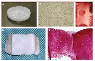
Figure 2: MSCs embedded in collagen hydrogel matrix used in treating cartilage defect in a pig. A) MSC embedded in a collagen matrix (obtained from a rat tail), B) A magnified view of MSC-containing collagen type 1 hydrogel matrix after 10 days of culture, C) Isolated chondral defect in trochlea of mini-pig, D) Macroscopic view of defect 6 months after treatment with MSCs, E) Immunohistochemical staining of the cartilage graft shows a cartilaginous, collagen type 11-rich extracellular matrix, which contains chondrocytes that are differentiated from MSCs [17].
Alternatively, MSCs are seeded into a biodegradable scaffold. This helps to support MSCs, especially in cases where the subchondral bone is exposed over large areas.
II Matrix-Guided Application of MSCs: The Way Forward
Compared with direct intra-articular MSC injection, matrix guided application of MSCs to cartilage offers better control and ease of application. Seeding or embedding of MSCs into a scaffold offers an accessible and easy way to manipulate self-renewable progenitor cells, which is in a very limited supply in the osteoarthritic joint. Numerous biomaterials have been tried as scaffolds, but none fulfills all the requirements. Ideally, a scaffold should be biodegradable, semi-permeable and adaptable to the mechanical environment, so as to promote the required activities of MSCs embedment in the joint.
In recent times, however, ‘natural’ scaffolds in the form of collagen type 1, chitosan and hyaluronan (hyaluronic acid) have been employed and they seem to present a more natural environment than synthetic scaffolds [16]. Collagen 11 hydrogels, for instance, offers three main advantages: firstly, they are biodegradable. Secondly, like hyaline cartilage, the collagen hydrogel in the osteoarthritic joint, can surround MSCs in a three-dimensional manner. In this way, the right environment is created to maximize the activity of MSCs inside the osteoarthritic joint. Lastly, hydrogels allow an even distribution of seeded-MSCs which helps to promote homogenous production of cartilage extracellular matrix within which the MSCs are embedded.
Direct intra-articular injection of MSCs has been well documented in animal models [18, 19]. There are now emerging data on the application of collagen hydrogel-seeded MSCs in human OA joints. In 2008, Schneider and colleagues evaluated the therapeutic benefit of CaReS®, a type 1 collagen hydrogel-based autologous chondrocyte implantation technique [20]. A total of 116 people with moderate to severe degree of knee OA treated via this technique had significant improvement in their symptoms.
However, the cost involved in constructing a scaffold from collagen has led to scientists looking into biomaterials with properties of collagen that can stimulate stem cell differentiation without growth factor supplementation. Singh et al. prepared a scaffold from cellulose and silk in a 75:25 ratio. After growing MSCs on it. They observed that chondrogenic marker genes SOX 9, aggrecan and type 11 collagen were upregulated in the absence of soluble growth factors such as tissue growth factor (TGF) β [21].
Conclusion
An insufficient response process of articular cartilage to inflammation contributes to the loss of phenotypic stability associated with osteoarthritic joints. Over the last two decades, numerous research studies have offered an insight into the science underpinning MSCs. In particular, we now know that MSCs proliferate and migrate into multiple skeletal tissues such as cartilage. The anti-inflammatory and immune-modulatory properties of MSCs have also been well documented, especially in the context of allogeneic transplantation. The hope is that MSCs will offer an alternative for OA treatment and patients will not need HLA matching and/or toxic medications. What is even more promising is that autologous and allogeneic MSC-based therapies for cartilage repair have been shown to produce acceptable clinical results in humans. Another major advance in MSC-based therapy is the discovery of new delivery modes for targeting MSCs into the joint.
The use of MSCs in combination with bioactive substrates; both natural and synthetic, has significant clinical potential and is likely to be important in future cartilage-repair technologies. In the long term, scientists and clinicians hope that MSC-based technologies will permit the engineering of cartilage not only for repair of focal lesions but also as a treatment option for large OA defects. More research is however required to realize the ultimate goal of a fully biological MSCs prosthesis and to translate their science into medicine.
Article Info
Article Type
Review ArticlePublication history
Received: Wed 09, Sep 2020Accepted: Sat 17, Oct 2020
Published: Fri 30, Oct 2020
Copyright
© 2023 J Agilinko. This is an open-access article distributed under the terms of the Creative Commons Attribution License, which permits unrestricted use, distribution, and reproduction in any medium, provided the original author and source are credited. Hosting by Science Repository.DOI: 10.31487/j.SCR.2020.10.04
Figures & Tables
References
- Fox AJS, Bedi A, Rodeo SA (2009) The Basic Science of Articular Cartilage: Structure, Composition, and Function. Sports Health 1: 461-468. [Crossref]
- Alford JW, Cole BJ (2005) Cartilage Restoration, Part 1: basic science, historical perspective, patient evaluation, and treatment options. Am J Sports Med 33: 295-306. [Crossref]
- Akkiraju H, Nohe A (2015) Role of Chondrocytes in Cartilage Formation, Progression of Osteoarthritis and Cartilage Regeneration. J Dev Biol 3: 177-192. [Crossref]
- Hwang HS, Kim HA (2015) Chondrocyte Apoptosis in the Pathogenesis of Osteoarthritis. Int J Mol Sci 16: 26035-26054. [Crossref]
- Sharif M, Whitehouse A, Sharman P, Perry M, Adams M (2004) Increased apoptosis in human osteoarthritic cartilage corresponds to reduced cell density and expression of caspase-3. Arthritis Rheumatism 50: 507-515. [Crossref]
- Gupta PK, Das AK, Chullikana A, Majumdar AS (2012) Mesenchymal stem cells for cartilage repair in osteoarthritis. Stem Cell Res Ther 3: 25. [Crossref]
- Minas T, Gomoll AH, Solhpour S, Rosenberger R, Probst C et al. (2009) Autologous Chondrocyte Implantation for Joint Preservation in Patients with Early Osteoarthritis. Clin Orthop Relat Res 468: 147-157. [Crossref]
- Yen YM, Cascio B, O’Brien L, Stalzer S, Millett PJ et al. (2008) Treatment of Osteoarthritis of the Knee with Microfracture and Rehabilitation. Med Sci Sports Exerc 40: 200-205. [Crossref]
- Dall’Oca C, Cengarle M, Costanzo A, Giannini N, Vacchiano A et al. (2017) Current concepts in treatment of early knee osteoarthritis and osteochondral lesions; the role of biological augmentations. Acta Biomed 88: 5-10. [Crossref]
- Mithoefer K, Williams RJ 3rd, Warren RF, Potter HG, Spock CR et al. (2006) Chondral resurfacing of articular cartilage defects in the knee with the microfracture technique. J Bone Joint Surg Am 88: 294-304. [Crossref]
- Dominici M, Blanc KL, Mueller I, Cortenbach IS, Marini F et al. (2006) Minimal criteria for defining multipotent mesenchymal stromal cells. The International Society for Cellular Therapy position statement. Cytotherapy 8: 315-317. [Crossref]
- Friedenstein AJ (1990) Osteogenic stem cells in bone marrow. In: Heersche JNM, Kanis JA, editors. Bone Mineral Res 243-272.
- Greene MA, Loeser RF (2015) Aging-related inflammation in osteoarthritis. Osteoarthritis Cartilage 23: 1966-1971. [Crossref]
- Barry F, Boynton RE, Liu B, Murphy JM (2001) Chondrogenic Differentiation of Mesenchymal Stem Cells from Bone Marrow: Differentiation-Dependent Gene Expression of Matrix Components. Exp Cell Res 268: 189-200. [Crossref]
- Weiss S, Hennig T, Bock R, Steck E, Richter W (2009) Impact of growth factors and PTHrP on early and late chondrogenic differentiation of human mesenchymal stem cells. J Cell Physiol 223: 84-93. [Crossref]
- Correia CR, Teixeira LSM, Moroni L, Reis RL, Blitterswijk CAV et al. (2011) Chitosan Scaffolds Containing Hyaluronic Acid for Cartilage Tissue Engineering. Tissue Eng Part C Methods 17: 717-730. [Crossref]
- Nöth U, Steinert, Tuan RS (2008) Technology Insight: adult mesenchymal stem cells for osteoarthritis therapy. Nat Clin Pract Rheumatol 4: 371-380. [Crossref]
- Sato M, Uchida K, Nakajima H, Miyazaki T, Guerrero AR et al. (2012) Direct transplantation of mesenchymal stem cells into the knee joints of Hartley strain guinea pigs with spontaneous osteoarthritis. Arthritis Res Ther 14: R31. [Crossref]
- Horie M, Sekiya I, Muneta T, Ichinose S, Matsumoto K et al. (2009) Intra-articular Injected Synovial Stem Cells Differentiate into Meniscal Cells Directly and Promote Meniscal Regeneration Without Mobilization to Distant Organs in Rat Massive Meniscal Defect. Stem Cells 27 878-887. [Crossref]
- Schneider U, Rackwitz L, Andereya S, Siebenlist S, Fensky F et al. (2011) A Prospective Multicenter Study on the Outcome of Type 1 Collagen Hydrogel–Based Autologous Chondrocyte Implantation (CaReS) for the Repair of Articular Cartilage Defects in the Knee. Am J Sports Med 39: 2558-2565. [Crossref]
- Singh N, Rahatekar SS, Koziol KKK, Ng TS, Patil AJ et al. (2013) Directing Chondrogenesis of Stem Cells with Specific Blends of Cellulose and Silk. Biomacromolecules 14: 1287-1298. [Crossref]