Journals
Thyroid-like follicular carcinoma of the kidney: A Case report
A B S T R A C T
Primary thyroid-like follicular carcinoma of the kidney (TLFCK) is a least-frequent and least-studied variant of renal tumor which is rich in follicular structures of colloidal substances, otherwise, it is typically negative for the thyroid immunohistochemical markers. We report a 44-year-old patient with mild anemia, who was found a right kidney occupying lesion incidentally during the medical examination. The microfollicular and macro-follicular structures under the microscope and typical negative performance of thyroid immunohistochemical markers is in line with the diagnosis of TLFCK. To our knowledge this is the first report of TLFCK with anemia. Brief review of previously published TLFCs, we discuss the clinical and histopathological features of limited cases.
K E Y W O R D S
Thyroid-like follicular carcinoma of the kidney, thyroid immunohistochemical markers, clinical and histopathological features
Introduction
TLFCK is a kind rare pathological type of renal tumor, which is potentially considered origining in the renal parenchymal urinary tubule epithelial system [1]. This unique histopathological type of RCC(renal cell carcinoma), described firstly in 1996 by Angell et al, had not been recorded in the 2004 world health organization in account of the lack of sufficient clinical evidence that only 28 cases reported in the English literature to date [2-15]. TLFCK has a similar feature with the follicular carcinoma of the thyroid in morphology. It is rich in follicular structures of colloidal substances, conversely, it is typically negative for the thyroid immunohistochemical markers [1]. Here we report a case of TLFCK with mild anemia and discuss the clinical presentation and histopathological features.
Case Report
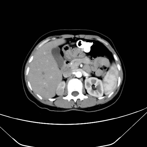
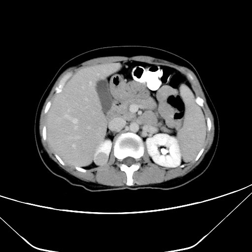
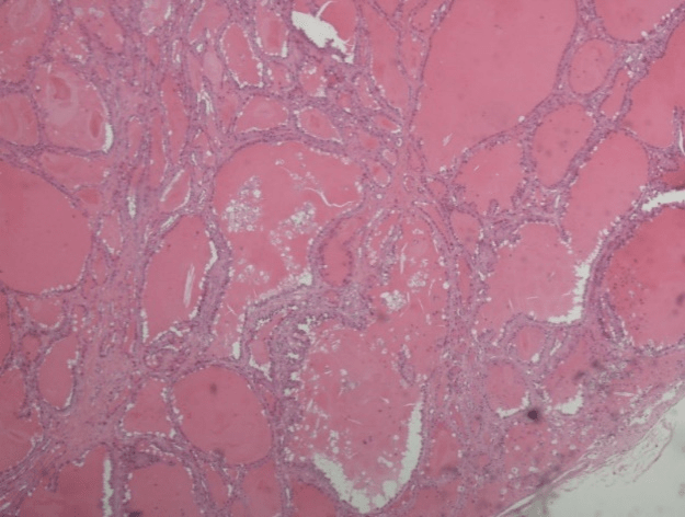
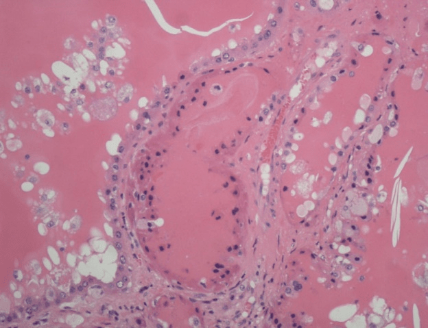
A 44-year-old woman who was found a right kidney occupying lesion incidentally during the physical examination was admitted to the hospital on April 21, 2017, no relevant history of related family disease noted. She had no gross hematuria, no flank pain, no weight loss, no obvious abnormality in the abdominal physical examination and no palpable lesion in the thyroid gland. Routine laboratory tests revealed that HGB was 87 g/L, HCT 32.10 %, MCV 63.2 fL. The results of the other routine laboratory tests, including blood routine, urine routine, liver and kidney function, blood biochemistry, coagulation series, PCT, were all within normal reference range. Electrocardiogram, chest radiography, lung function test was all negative. ECT prompted a delay of the right renal pelvis drainage. CFR suggested that bilateral renal function was normal. Computer tomography (CT) demonstrated that a heterogeneous mass with obvious calcification and well-defined border, low contrast enhancement, measuring 20mm, in the right kidney (Figure 1). There was no evidence indicated the visible invasion of the renal vascular and regional enlarged lymph nodes. The patient underwent a laparoscopic partial right hepatectomy. Microscopically, the carcinoma is composed of microfollicular and macro-follicular structures similar to thyroid follicles (Figure 2). Immunohistochemically, the tumor was positive for CK7, and negative for TG, TTF1, CD10, P504S, CD117 (Figure 3).
Discussion
Primary thyroid-like follicular carcinoma of the kidney(TLFCK) is a least-frequent and least-studied variant of renal tumor, named purely from the light microscopic appearance, with well-circumscribed pseudo-capsules resembling to thyroid follicular carcinoma microscopically but negative for the thyroid-specific markers [2, 3, 15]. In all reported cases, there were no significant distinguish from other types of renal tumor on location and clinical performance. The age of most patients ranges 19 to 83 years old, and the female is more effected than the male [1, 5, 11, 12]. Most of them were incidentally found with or without other clinically positive performance [14]. The macroscopic hematuria and abdominal pain are thought as the common characteristics around TLFCK [10]. It is also difficult to distinguish between TLFCK and other pathological types of renal cancer throgh imaging performance, as there has been no specific clinical characteristics of TLFCK. In particular, the patient, in this case, was suffering from mild anemia, and the condition gradually recovers after surgery. But, the association between TLFCK and anemia remains unclear.
The diagnosis of TLFCK mainly depends on the microscopic appearance and immunohistochemical experience. Morphologically, TLFCK consists of different sized follicular structures containing abundant eosinophilic colloid resembling to the well-differentiated follicular thyroid carcinoma, as a comparison, TLFCK show a obviously different morphological features from other types of renal carcinoma[6]. Immunohistocemically, TLFCK has a typical negative performance of thyroid immunohistochemical markers, like thyroid transcription factor-1(TTF1) and thyroglobulin(TG), respectively, the staining of CD10 and CK17 is variable [1, 3, 5, 7]. In this case, Immunohistochemical results show a positive result of CK7, and negative result of TTF1, TG, P504S, CD117, meeting the diagnostic criteria of TLFCK.
The differential diagnosis of TLFCK includes renal metastases of thyroid carcinoma and thyroidization of renal tubules. Thyroidization of renal tubules is a well-known phenomenon, of which the general structure of atrophic distal tubules or collective tubules is similar to that of colloidally transparent tubes of thyroid [14]. In general, the process of glandularization occurs after the chronic pyelonephritis or obstructive nephropathy or the end-stage renal disease [7]. Whereas, TLFCK, which appears as a clear-cut mass, occurs in the patients without suffering from kidney disease attack. TLFCK is composed almost exclusively of dense follicular structures and colloidal substances. The renal tumors described herein are histologically similar to follicular thyroid cancer but still should be distinguished from metastatic thyroid cancer. Thyroid follicular carcinoma usually metastasizes to the lungs and bone, rarely to the kidney. In addition, the tumor shows a positive reaction of thyroid-specific markers TTF-1 and TG [8]. The available data indicated that TLFRCC immunohistochemical staining shows a consistent expression of the transcription factor PAX8, and a typical negative performance of thyroid immunohistochemical markers, which is significant to the identification of thyroid metastatic tumors. Therefore, on the basis of morphology and the result of f immunohistochemistry, the diagnosis of this tumor as TLFCK was made.
The surgery is still the referred treatment [11]. Among the reported TLFCK cases, all of them were low grade and indolent ,with only two cases metastases, and no reports of death related to the disease [10].

Disclosure
This study was supported by the Key Project of Research and Development Plan in Shandong Province (No.2016GSF201172) the Scientifc Research Foundation of Shandong Province for Outstanding Young Scientist Award (No. BS2014YY036) the National Natural Science Foundation of China (No. 81472395 and NO.81672522). b
Article Info
Article Type
Case ReportArticle History
Received 13 October, 2018Accepted 2 November, 2018
Published 29 November, 2018
Copyright
© 2018 The Authors. This is an open-access article distributed under the terms of the Creative Commons Attribution License, which permits unrestricted use, distribution, and reproduction in any medium, provided the original author and source are credited. Hosting by Science Repository.10.31487/j.SCR.2018.03.015
Author Info
Corresponding author
Yidong FanDepartment of Urology, Qilu Hospital of Shandong University, Jinan 250012, China.
Zhiqing Fang
Department of Urology, Qilu Hospital of Shandong University, Jinan 250012, China.
Figures & Tables
References
1. Wu WW, Chu JT, Nael A, Rezk SA, Romansky SG, et al. (2014) Thyroid like follicular carcinoma of the kidney in a young patient with history of pediatric acute lymphoblastic leukemia. Case Rep Pathol 2014: 313974. [Crossref]
2. Cao D (2013) Thyroid-like follicular carcinoma of the kidney. J Urol 190: 278-279.
3. Alexandre Cavalcante, André Y. Kuwano, André Costa-Matos, Ezequiel F. Spanholi, Túlio Souza, et al. (2017) Thyroid-like follicular carcinoma of the kidney - Case report. Urol Case Rep 15: 36-38. [Crossref]
4. Chen F, Wang Y, Wu X, Zhu Y, Jiang X, et al. (2016) Clinical characteristics and pathology of thyroid-like follicular carcinoma of the kidney: Report of 3 cases and a literature review. Mol Clin Oncol 4: 143-150. [Crossref]
5. Dawane R, Grindstaff A, Parwani AV, Brock T, White WM, et al. (2015) Thyroid-like follicular carcinoma of the kidney: one case report and review of the literature. Am J Clin Pathol 144: 796-804. [Crossref]
6. Dhillon J, Mohanty SK, Krishnamurthy S (2014) Cytologic diagnosis of thyroid-like follicular carcinoma of the kidney: a case report. Diagn Cytopathol 42: 273-277. [Crossref]
7. Jasreman Dhillon, Nizar M. Tannir, Surena F. Matin, Pheroze Tamboli, Bogdan A. Czerniak, et al. (2011) Thyroid-like follicular carcinoma of the kidney with metastases to the lungs and retroperitoneal lymph nodes. Hum Pathol 42: 146-150. [Crossref]
8. Dong L, Huang J, Huang L, Shi O, Liu Q, et al. (2016) Thyroid-Like Follicular Carcinoma of the Kidney in a Patient with Skull and Meningeal Metastasis: A Unique Case Report and Review of the Literature. Medicine (Baltimore) 95: 3314. [Crossref]
9. Ghaouti M, Roquet L, Baron M, Pfister C, Sabourin JC (2014) Thyroid-like follicular carcinoma of the kidney: a case report and review of the literature. Diagn Pathol 9: 186. [Crossref]
10. Kryvenko ON, Jorda M, Argani P, Epstein JI (2014) Diagnostic approach to eosinophilic renal neoplasms. Arch Pathol Lab Med 138: 1531-1541. [Crossref]
11. Lee JG, Yang Y, Kim KS, Hyun CL, Lee JS, et al. (2011) A case of metastatic renal cell carcinoma to thyroid gland. Chonnam Med J 47: 130-133. [Crossref]
12. Li C, Dong H, Fu W, Qi M, Han B et al. (2015) Thyroid-like Follicular Carcinoma of the Kidney and Papillary Renal Cell Carcinoma with Thyroid-like Feature: Comparison of Two Cases and Literature Review. Ann Clin Lab Sci 45: 707-712. [Crossref]
13. Lin YZ, Wei Y, Xu N, Li XD, Xue XY, et al. (2014) Thyroid-like follicular carcinoma of the kidney: A report of two cases and literature review. Oncol Lett 7: 1796-1802. [Crossref]
14. Malde S, Sheikh I, Woodman I, Fish D, Bilagi P, et al. (2013) Primary thyroid-like follicular renal cell carcinoma: an emerging entity. Case Rep Pathol 2013: 687427. [Crossref]
15. Nath V, et al. (2015) Follicular Thyroid Carcinoma Metastatic to the Kidney: Report of a Case with Cytohistologic Correlation. Case Rep Pathol 2015: 701413.