
The Role of the Surgery in the Case of an Intramedullary Spinal Cord Metastasis as a Primary Presentation of a Malignant Disease
A B S T R A C T
Introduction: Spinal cord metastases represent a small proportion of intramedullary tumors. The occurrence of such lesion over the course of malignant disease is usually a predictor for shortened life expectancy and is often associated with severe neurological deficits. Treatment options include microsurgical excision, radio-, chemo- or palliative therapy. Despite these possibilities the optimal management of patients with intramedullary spinal cord metastases (ISCM) is difficult due to the wide variety of clinical situations and the lack of controlled studies on the results of different therapeutic options.
Materials: We are presenting a case of a 68-year-old male with gradually increasing spinal neurological deficit – axial low back pain, numbness and muscle weakness in both legs and gait disturbance. Arterial hypertension was pointed as the only comorbidity of the patient. The MRI showed an intramedullary solitary lesion in the lumbar intumescence at the level of Th12 and L1 vertebra.
Results: The patient underwent surgery and the tumor was totally excised. The symptoms improved dramatically. The histology evidence a metastasis from a lung adenocarcinoma. Six months later he presented with severe back pain, progression of the paresis and urinary retention. Tumour recurrence was detected by MRI. After careful decision making and taking into consideration the risk of permanent neurological deficit a reoperation was performed. The lesion was gross-totally removed with improvement in functions and no neurological deterioration registered in the postoperative period.
Conclusion: It is possibly the symptoms from an intramedullary metastatic lesion to precede the detection of the primary tumour. The low frequency of occurrence and the absence of a manifested and diagnosed primary malignant process should not stop us thinking in the direction of metastasis. Surgical resection with function preservation is highly aimed. Operation and even reoperation in some cases is acceptable.
Keywords
Intramedullary tumor, spinal cord metastasis, lung cancer, surgery
Introduction
Spinal cord metastases represent a small proportion of intramedullary tumors. They are estimated to affect between 0.5% and 2.0% of all patients with cancer, rep¬resenting only 8.5% of all CNS metastases and compris¬ing 1–3% of all intramedullary spinal cord tumours (ISCT) [1-4]. Compared with neoplastic epidural compression, intramedullary spinal cord metastasis (ISCM) are diagnosed much less frequently, with an incidence approximately one-sixteenth as high [1, 2, 5]. The occurrence of such pathology over the course of malignant disease is usually a predictor for shortened life expectancy and is often associated with severe neurological deficits. It can also sug¬gests an advanced form of cancer and a biologically aggressive disease.
ISCT can arise as metastatic deposits within the spinal cord parenchyma from any type of primary cancer, but close to 50% of them result from primary lung cancer, with small cell carcinoma being the most common, and the remainder originating from primary cancers of the breast, colon, kidney, from melanomas and lymphomas [6]. The most frequent pathway for dissemination is the arterial route, direct invasion through the spinal roots or leptomeninges can also occur and retrograde spread through the Batson plexus during the Valsava maneuver has also been suggested [7]. Based on the data in the literature the cervical segment of the cord is the most and the lumbar is the least affected, with thoracic segment staying in between [8-10]. Symptoms at presentation are usually pain and/or weakness. They are often followed by sensory level and sphincter disturbance. Rapid deterioration, within a period of days to weeks, with progression to a cord hemisection syndrome (Brown-Sequard) or full cord transection is possible.
Case Description
A 68-year-old male presenting with a signs of spinal cord compression - axial low back pain, numbness and muscle weakness in both legs and gait disturbance. The symptoms appeared 10 days ago and increased gradually in time. Arterial hypertension was pointed as the only comorbidity of the patient. The neurological examination revealed mild flaccid paresis, decreased reflexes and hypesthesia in the lower limbs. The patient underwent MRI of the spine, showing single solitary intramedullary lesion, located in the lumbar intumescence at the level of Th12 and L1 vertebrae (Figure 1). The lack of history for primary cancer made us assume that the type of the tumour is an ependymoma. This and the progression of the neurological symptoms were perceived as indications for surgery.

Figure 1: Sagittal and axial contrast-enhanced MRI images, showing intramedullary tumour at the level of Th12 and L1 vertebrae.
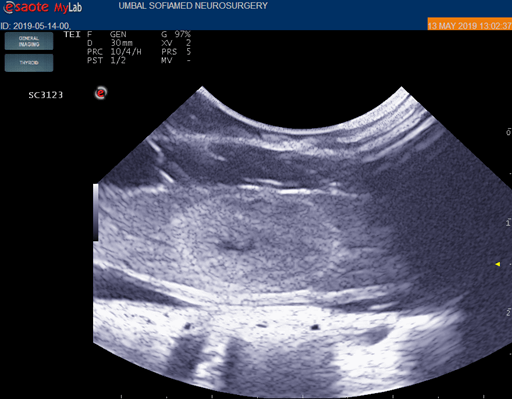
Figure 2: Intraoperative ultrasound view of the tumour.
A standard operative approach was used – prone position, median skin incision, bilateral paraspinal muscle stripping, laminectomy of Th12 and L1 vertebrae. Then ultrasound was used for verifying the exact location of the lesion (Figure 2), followed by opening the dura (Figure 3) and small myelotomy right over the tumor projection (Figure 4). After careful dissection the tumor was gross totally removed (Figure 5) with no changes in the intraoperative monitoring parameters. No new neurological deficit was observed after the surgery. The muscle strength increase, and the pain diminish in the postoperative period, the sensory disturbance remains unchanged. The histological diagnosis, including immunohistochemical analysis substantiate non-small-cell lung adenocarcinoma. The patient was discharged and referred for subsequent treatment in oncology department. Three months after the surgery he underwent spine MRI for disease follow up. There were no symptoms present, however a small tumor relapse [with a diameter 0,85 cm] was evidenced (Figure 6).
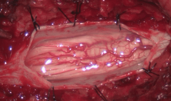
Figure 3: Intraoperative image after opening the dura, showing the spinal cord at the level of the lumbar intumescence.
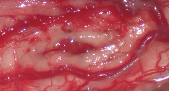
Figure 4: Intraoperative image showing the site of the myelotomy. The incision is located between the pial veins of the coronal venous plexus.
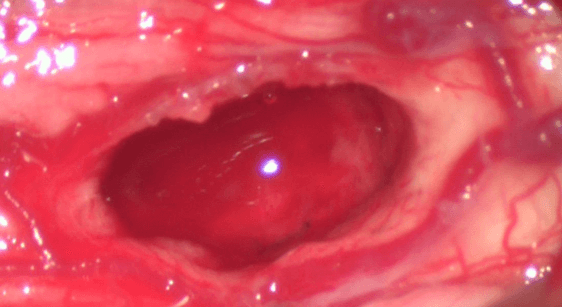
Figure 5: Intraoperative image after removal of the lesion, showing the tumor cavity in the spinal cord.
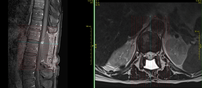
Figure 6: Sagittal and axial MRI images, performed 3 months after the surgery, showing small relapse of the lesion.
Operative treatment was offered, but declined, mainly due to the lack of clinical manifestation. The prime treatment method was chemotherapy and it was relied on its effect. After another three months or totally six after the surgery the patient was admitted with severe back pain, paraparesis, causing inability of standing and walking and newly emerged urine retention. The conducted MRI showed distinct increase in tumor’s size (Figures 7 & 8). After careful discussion, taking into account the patient’s wish for carrying out the surgery, and on the other hand, the potential risk for permanent neurological morbidity, a decision for performing a reoperation was made. The same surgical approach was undertaken, a lot of adhesions and a retained epidural CSF collection were present. The lesion was located mainly in the tumor cavity, which made possible it’s complete removal. The patient recovered well, the paraparesis improved, the pain was gone and there was no evidence for urine retention. He was discharged, fully able to walk on his own and referred to oncology for continuing the treatment.
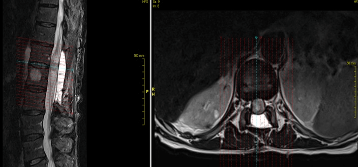
Figure 7: Sagittal and axial MRI images, performed 6 months after the surgery, showing clear increase in the size of the lesion.
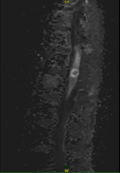
Figure 8: Sagittal diffusion-weighted MRI, performed 6 months after the surgery, showing clear increase in the size of the lesion.
Discussion
The proper diagnosis of ISCT is essential to provide the best treatment and obtain the optimal outcome. Intramedullary secondary lesions usually occur in the con¬text of a well-established diagnosis of cancer. However, in some patients, the manifestation of symptoms can be misleading, or the primary disease can be occult and then the diagnosis can be difficult. Sung et al. performed a retrospective review of 301 patients and found that in 26% of the cases the detection of the ISCM led to the ultimate diagnosis of the systemic cancer [1]. By the majority of these patients the definitive histological diagnosis of the ISCM and the primary disease resulted from a surgical biopsy or resection [11-23].
There are some signs that can suggest a secondary lesion. While slow progression of the symptoms (mainly mild pain and neurological deficit) is typical for primary spinal cord tumours, one rapid deterioration speaks for a metastatic lesion. In addition, muscular atrophy and fasciculation are being more frequently observed in primary intramedullary lesions when compared with ISCM [24, 25]. On the other hand, MRI can be very helpful – the metastases are usually solid with no cystic or haemorrhagic components (which can be present in the case of primary tumour) and there is distinct contrast enhancement with the so-called ,,rim’’ and ,,flame’’ signs. The first is a complete or partial, thin, peripheral rim of gadolinium enhancement more intense than the central enhancement of a noncystic/necrotic lesion and the second is an ill-defined flame-shaped region of gadolinium enhancement at the superior and/or inferior margin of an otherwise well-defined lesion [26].
And why a precise preoperative diagnosis is so important? Because of that depends the type of subsequent approach. The treatment modalities include surgery, radiation therapy (RT), chemotherapy (CT) or steroids. Surgery is the preferred choice in the case of primary intramedullary lesion. The goal is gross total removal, insofar as possible, without the occurrence of new neurological deficit. In the case of ependymoma (the most common spinal cord tumour in adults) or low-grade astrocytoma that would lead to cure of the disease, because relapse wouldn’t occur or that could happen extremely rare. When it comes to high-grade astrocytoma or some other more malignant version of the primary tumour, where the lesion is infiltrative and borders with normal tissues are lost, the purpose of surgical treatment is to decompress the spinal cord and by that to prevent the progression of clinical symptoms and the onset of a persistent neurological mortality.
In the case of metastasis things are different. RT has been the treatment of choice since many years. Best responses are achieved in radiosensitive cases especially if implemented very early over the course of presentation [4]. Because of the risk for developing radiation myelitis a fractionated radiotherapy (FR) or stereotactic radiosurgery (SRS) are recently recommended. CT has been used in conjunction with RT or surgery in some chemotherapy-sensitive tumours. According to some authors, CT is not effective in the case of ISCM, because the drugs do not pass through the blood-spinal cord barrier (BSB) and have no further effect on survival [27-30].
Patients with rapidly progressive symptoms of cord compression and a high risk of rapid deterioration are suitable for treatment with high-dose steroids. This may decrease the pain and cause transient improvement in neurological conditions. Steroids suppress perifocal oedema and normalize the BSB, which decreases tumour size but does not influence survival [7].
The most common indications for performing surgery are the decompression of functional neural tissue and histological confirmation of the tumour. Currently, surgical approach is more precise and less invasive and thus allows total resection with no or acceptable morbidity rate. Considering the importance of total tumour resection in the case of cerebral metastases, one could postulate a similar importance when surgically removing ISCM. Kalayci et al. demonstrated the length of survival in surgical patients being almost twice as long than the nonsurgical patients [4]. Connolly et al. also found that the survival correlated strongly with aggressiveness of therapy [12]. However, Wilson et al. as well as Watanabe et al. could not find a benefit from surgery concerning the length of survival in their respective series [23, 31]. Nevertheless, their patients had preoperative urinary retention, which is accompanied by a progressive neurological deficit and poor prognosis. Our patient also had that symptom before the second surgery.
Performing a reoperation for an intramedullary lesion is always a hazardous decision. The data in the literature reporting such action is very limited. Our considerations included the explicit wish of the patient for surgical intervention. He had severe pain and couldn’t bear it. Even though the relapse of the lesion was bigger in size, there were less tumour blood vessels present and less bleeding, which made the dissection easier. Working only within the boundaries of the tumour, using careful microdissection technique, permitted gross total removal with no changes in the neurophysiological parameters and no appearance of neurological morbidity after the surgery.
Conclusion
It is possibly the symptoms from an intramedullary metastatic lesion to precede the detection of the primary tumour. The low frequency of occurrence and the absence of a manifested and diagnosed primary malignant process should not stop us thinking in the direction of metastasis. Surgical resection with function preservation is highly aimed. Operation and even reoperation in some cases is acceptable and can potentially result in significant improvement in the neurological function and quality of life.
Abbreviation
BSB: Blood-spinal cord barrier
ISCM: Intramedullary spinal cord metastasis
ISCT: Intramedullary spinal cord tumours
CNS: Central nervous system
CT: Chemotherapy
RF: Fractionated radiotherapy
RT: Radiation therapy
SRS: Stereotactic radiosurgery
Article Info
Article Type
Case ReportPublication history
Received: Thu 19, Mar 2020Accepted: Tue 07, Apr 2020
Published: Mon 13, Apr 2020
Copyright
© 2023 S. Byulbyuleva. This is an open-access article distributed under the terms of the Creative Commons Attribution License, which permits unrestricted use, distribution, and reproduction in any medium, provided the original author and source are credited. Hosting by Science Repository.DOI: 10.31487/j.SCR.2020.04.01
Figures & Tables
References
- Sung WS, Sung MJ, Chan JH, Manion B, Song J et al. (2013) Intramedullary spinal cord metastases: a 20-year institutional experience with a comprehensive literature review. World Neurosurg 79: 576-584. [Crossref]
- Rykken JB, Diehn FE, Hunt CH, Schwartz KM, Eckel LJ et al. (2013) Intramedullary spinal cord metastases: MRI and relevant clinical features from a 13-year institutional case series. AJNR Am J Neuroradiol 34: 2043-2049. [Crossref]
- Chi JH, Parsa AT (2006) Intramedullary spinal cord metastasis: clinical management and surgical considerations. Neurosurg Clin N Am 17: 45-50. [Crossref]
- Kalayci M, Cagavi F, Gul S, Yenidünya S, Açikgöz B (2004) Intramedullary spinal cord metastases: diagnosis and treatment- an illustrated review. Acta Neurochir (Wien) 146: 1347-1354. [Crossref]
- Newton HB (1999) Neurological complications of systemic cancer. Am Fam Phys 59: 878-886. [Crossref]
- Schiff D, O’Neill BP (1996) Intramedullary spinal cord metastases: clinical features and treatment outcome. Neurology 47: 906-912. [Crossref]
- Kalita O (2011) Current insights into surgery for intramedullary spinal cord metastases: a literature review. Int J Surg Oncol 2011: 989506. [Crossref]
- Findlay JM, Berstein M, Vanderlinden RG, Resch L (1987) Microsurgical resection of solitary intramedullary spinal cord metastases. Neurosurgery 21: 911-915. [Crossref]
- Potti A, Abdel-Raheem M, Levitt R, Schell DA, Mehdi SA (2001) Intramedullary spinal cord metastases (ISCM) and non-small cell lung carcinoma (NSCLC): clinical patterns, diagnosis and therapeutic considerations. Lung Cancer 31: 319-323. [Crossref]
- Tognetti F, Lanzino G, Calbucci F (1988) Metastases of the spinal cord from remote neoplasms. Surg Neurol 30: 220-227. [Crossref]
- Aryan HE, Farin A, Nakaji P, Imbesi SG, Abshire BB (2004) Intramedullary spinal cord metastasis of lung adenocarcinoma presenting as Brown-Sequard syndrome. Surg Neurol 61:72-76. [Crossref]
- Connolly ES Jr, Winfree CJ, McCormick PC, Cruz M, Stein BM (1996) Intramedullary spinal cord metastasis: report of three cases and review of the literature. Surg Neurol 46: 329-337; discussion 337-328. [Crossref]
- Cordan T, Bekar A, Tureyen K, Dogan S, Tolunay S (1994) Intramedullary spinal cord metastasis: case report. Turkish Neurosurgery 4: 171-173.
- Denaro L, Pallini R, Di Muro L, Ciampini A, Vellone V et al. (2007) Primary hemorrhagic intramedullary melanoma. Case report with emphasis on the difficult preoperative diagnosis. J Neurosurg Sci 51: 181-183. [Crossref]
- Findlay JM, Bernstein M, Vanderlinden RG, Resch L (1987) Microsurgical resection of solitary intramedullary spinal cord metastases. Neurosurgery 21: 911-915. [Crossref]
- Grasso G, Meli F, Patti R, Giambartino F, Florena AM, Lacopino DG (2007) Intramedullary spinal cord tumor presenting as the initial manifestation of metastatic colon cancer: case report and review of the literature. Spinal Cord 45: 793-796. [Crossref]
- Guppy KH, Wagner F (2006) Metastasis to the conus medullaris: case report. Neurosurgery 59: E1148. [Crossref]
- Maiuri F, Giamundo A, Gangemi M (1986) Intramedullary spinal cord metastasis. A case report. Acta Neurol (Napoli) 8: 487-490.
- Marquart C, Weckesser M, Schueller P, Hasselblatt M, Wassmann H et al. (2007) Intramedullary spinal cord metastasis as initial presentation of systemic cancer—report of a rare case. Zentralbl Neurochir 68: 214-216. [Crossref]
- Schijns OE, Kurt E, Wessels P, Luijckx GJ, Beuls EA (2000) Intramedullary spinal cord metastasis as a first manifestation of a renal cell carcinoma: report of a case and review of the literature. Clin Neurol Neurosurg 102: 249-254. [Crossref]
- Sutter B, Arthur A, Laurent J, Chadduck J, Friehs G et al. (1998) Treatment options and time course for intramedullary spinal cord metastasis. Report of three cases and review of the literature. Neurosurg Focus 4: e3. [Crossref]
- Tognetti F, Lanzino G, Calbucci F (1988) Metastases of the spinal cord from remote neoplasms. Study of five cases. Surg Neurol 30: 220-227. [Crossref]
- Watanabe M, Nomura T, Toh E, Sato M, Mochida J (2006) Intramedullary spinal cord metastasis: a clinical and imaging study of seven patients. J Spinal Disord Tech 19: 43-47. [Crossref]
- Grem JL, Burgess J, Trump DL (1985) Clinical features and natural history of intramedullary spinal cord metastasis. Cancer 56: 2305-2314. [Crossref]
- Jellinger K, Kothbauer P, Sunder-Plassmann E, Weiss R (1979) Intramedullary spinal cord metastases. J Neurol 220: 31-41.
- Rykken B, Diehn FE, Hunt CH, Eckel LJ, Schwartz KM et al. (2013) Rim and Flame Signs: Postgadolinium MRI Findings Specific for Non-CNS Intramedullary Spinal Cord Metastases. Am J Neuroradiol 34: 908-915.
- Kosmas C, Koumpou M, Nikolaou M, Katselis J, Soukouli G et al. (2005) Intramedullary spinal cord metastases in breast cancer: report of four cases and review of the literature. J Neurooncol 71: 67-72. [Crossref]
- Isoya E, Saruhash Y, Katsuura A,Takahashi S, Matsusue Y et al. (2004) Intramedullary spinal cord metastasis of ovarian tumor. Spinal Cord 42: 485-487. [Crossref]
- Kodama M, Kawaguchi H, Komoto Y, Takemura M (2010) Coexistent intramedullary spinal cord and choroidal metastases in ovarian cancer. J Obstet Gynaecol Res 36: 199-203. [Crossref]
- Choi HC, Yoon DH, Kim SC, Cho KH, Kim SH (2010) Two separate episodes of intramedullary spinal cord metastasis in a single patient with breast cancer. J Korean Neurosurg Soc 48: 162-165. [Crossref]
- Wilson DA, Fusco DJ, Uschold TD, Spetzler RF, Chang SW (2012) Survival and functional outcome after surgical resection of intramedullary spinal cord metastases. World Neurosurg 77: 370-374. [Crossref]