
Reconstruction of a Wide Defect in the Prostatic Urethra after Abdomino-Perineal Amputation Using Buccal Mucosa Free Graft and Pedicled Gracilis Muscle Flap
A B S T R A C T
Lesions with significant substance loss in the prostatic segment of the urethra can represent a reconstructive challenge, especially when peripheral tissues are damaged or poor-vascularized. We present an infrequent clinical case in which a prostatic urethral defect of 2.6cm in length following abdomino-perineal amputation was repaired using a free buccal mucosa graft which was stabilized with a muscular gracilis flap. Complementing the buccal mucosa graft with a well-vascularised support the stability of the graft could be enhanced and the rates of fistula or strictures reduced.
Keywords
Urethral reconstruction, gracilis muscle, buccal mucosa, abdomino-perineal amputation, fistulae
Introduction
Lesions in the prostatic urethra are much less frequent than those in the bulbar or membranous urethra. The main aetiology of damage in the posterior urethra are pelvic fractures, but in these cases most of the lesions are located in the membranous or bulb membranous segments [1]. Restoration of urethral continuity through direct anastomosis is the technique of choice when possible. When a direct anastomosis is not possible due to urethral substance loss, it is necessary to apply reconstructive methods using local or distant tissue [2].
Case Report
Male 57-years-old presenting locally advanced rectal carcinoma (T3N1M0). Neoadjuvant radiotherapy and chemotherapy were administered prior to a laparoscopic Millie’s abdomino-perineal amputation. A tumour in the lower third of the rectum, firmly adhered to the prostatic capsule, was removed along with the posterior prostate lobe. Due to the extension of the perineal defect, a Mikulicz drainage was placed in the wound. The urinary catheter was removed 8 days after surgery and spontaneous micturition was observed. In successive days the gauzes of the Mikulicz drainage were progressively removed without incident but when the last gauze was removed, there was spontaneous urine drainage through the perineal wound. Physical examination revealed a defect of 2.6 cm in length in the posterior wall of the prostatic urethra. Reconstructive surgery was programmed three days later.
The patient was placed under general anaesthesia in an exaggerated lithotomy position. A suprapubic cystostomy was performed in order to guarantee bladder urine drainage. A 3.0cmx1.0cm buccal mucosa graft was harvested from the vestibule of the lower lip; the wound in the mouth was primarily closed. In the perineal region, dissection of the margins of the urethral defect was carried out and they were adapted and sutured to the buccal mucosa graft using the bladder catheter as an internal tutor (Figure 1).
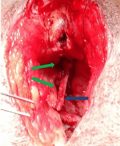
Figure 1: perineal wound with the urethral defect (green arrows) and the buccal mucosa graft partially sutured (blue arrow).
With the aim of providing the buccal mucosa graft with a stable and well-vascularised support, we decided to harvest a gracilis muscle flap (GM-F) based on its main vascular pedicle from the left thigh. The pedicle of the flap was entirely dissected to its origin in the medial femoral circumflex artery and the muscle was transected from its distal insertions. The flap was folded back through a skin incision to the perineal wound (Figure 2A) and the distal portion of the gracilis muscle was sutured at the base of the perineal wound providing a meticulous adaptation of the muscular flap surface over the buccal mucosa graft thus avoiding any space between the two surfaces (Figure 2B). A vacuum drain was inserted, and the GM-F was anchored to the walls of the wound. The perineal skin was primarily closed as in the donor area of the GM-F. The patient was confined to bed for six days following the operation. No adverse event was registered during the immediate post-operative period and the patient was discharged 23 days after surgery with both a cystostomy and a bladder catheter.
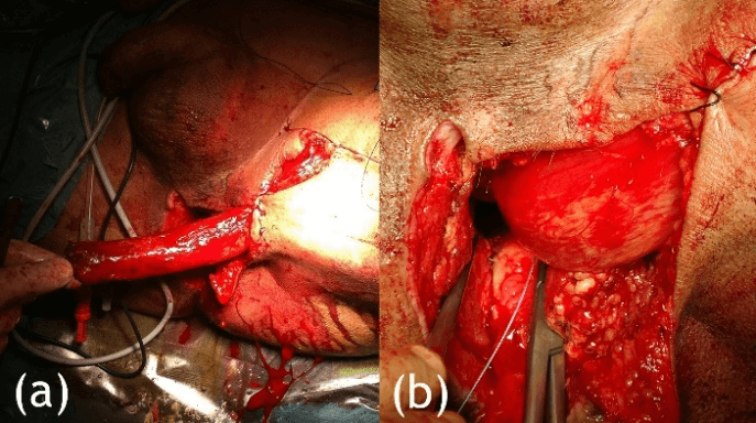
Figure 2: A) the left gracilis muscle is harvested from its main pedicle and folded back to reach easily the perineal defect. B) the distal end of the gracilis muscle flap is sutured to the base of the perineal wound covering the urethral buccal mucosa graft.
The bladder catheter was removed a month and a half after surgery and normal micturition was observed. A cystography revealed the integrity of the reconstructed urethra with no stricture segment or fistulae. The cystostomy catheter was removed a week later. Eleven months after the urethral reconstruction, the patient does not present any complications. A cystography has revealed a successful integration of the buccal mucosa graft and no fistula or stricture have been noted.
Discussion
It is clear that there is not as much clinical experience in the reconstruction of the posterior urethral segment as there is in the case of the anterior segment. Non-glabrous full-thickness free skin grafts have presented a great number of complications including urethral stricture in the operated area [3]. Split-thickness skin grafts seem to present a lesser tendency towards stricture but with the disadvantage that the elasticity and resistance properties of the graft are reduced [4]. Bladder mucosa grafts have gained in popularity in recent years, but a more aggressive abdominal approach is necessary to obtain them, and they are not exempt from post-operatory complications [5]. In our experience buccal mucosa grafts are the most appropriate option to restore a urethral defect. The harvesting of the graft is a simple process and the donor site hardly ever presents any significant post-operative complications. Indeed, some articles have shown that buccal mucosa grafts present a post-operative complication rate lower than 10% in the grafted area [6].
The gracilis flap, harvested as a muscle flap or combined with a fat and skin paddle, has been widely used in perineal and genital area coverture [7-9]. In the case presented here, the poor vascularisation of the local tissues made it necessary to supply healthy and well-vascularised distant tissues in order to allow a suitable wound coverture and a stable support to the buccal mucosa graft. Furthermore, the volume provided by the muscular flap was employed to occupy the empty space in the perineal wound. This allowed the direct closure of the wound reducing the total healing time.
To cover abdomino-perineal amputation defects some authors have made use of rectus abdominis muscle flaps with low rates of post-operative complications, even when patients had undergone radiotherapy in the operated area [10]. In this particular case, we decided that a rectus abdominis muscle flap would produce higher morbidity without any advantage for the patient. We strongly believe that any damage in the abdominal wall should be avoided if possible and a GM-F allows good coverture without any considerable functional damage. Although long term evolution is still to be assessed we think that complementing the buccal mucosa graft with a well-vascularised support could enhance the stability of the graft and reduce the rates of fistula or strictures.
Conclusion
Although urethral lesions with substance loss in the prostatic segment that could not be primarily repaired are infrequent in abdomino-perineal amputation, they could represent a complication with a significant degree of morbidity in a patient that will undergo adjuvant therapy after surgery. An early restoration of the urethral continuity using a buccal mucosa graft complemented with a GM-F represents a simple and reliable solution. This allows an early recovery, thus making a better continuation of the oncological adjuvant treatment possible.
Conflicts of Interest
None.
Article Info
Article Type
Case ReportPublication history
Received: Tue 12, May 2020Accepted: Mon 25, May 2020
Published: Mon 01, Jun 2020
Copyright
© 2023 Ivan Couto González. This is an open-access article distributed under the terms of the Creative Commons Attribution License, which permits unrestricted use, distribution, and reproduction in any medium, provided the original author and source are credited. Hosting by Science Repository.DOI: 10.31487/j.SCR.2020.06.02
Figures & Tables
References
- Jordan GH, Virasoro R, Eltahamy EA (2006) Reconstruction and Management of Posterior Urethral and Straddle Injuries of the Urethra. Urol Clin N Am 3: 97-109. [Crossref]
- Secrest CL (2002) Staged Urethroplasty: Indications and Techniques. Urol Clin N Am 29: 467-475. [Crossref]
- Markiewicz MR, Lukose MA, Margarone JE 3rd, Barbagli G, Miller KS et al. (2007) The Oral Mucosa Graft: A Systematic Review. J Urol 178: 387-394. [Crossref]
- Kröpfl D, Tucak A, Prlic D, Verweyen A (1998) Using Buccal Mucosa for Urethral Reconstruction in Primary and Re-Operative Surgery. Eur Urol 34: 216-220. [Crossref]
- Watanabe K, Ogawa A, Kiyono M (1995) Reconstruction of the Urethral Meatus With a Buccal Mucosa Graft. Urol Int 55: 29-31. [Crossref]
- Elliott SP, Metro MJ, McAninch JW (2003) Long-term Followup of the Ventrally Placed Buccal Mucosa Onlay Graft in Bulbar Urethral Reconstruction. J Urol 169: 1754-1757. [Crossref]
- Vyas RM, Pomohac B (2010) Use of a Bilobed Gracilis Myocutaneous Flap in Perineal and Genital Reconstruction. Ann Plast Surg 65: 225-227. [Crossref]
- Chen SY, Fu JP, Chen TM, Chen SG (2011) Reconstruction of Scrotal and Perineal Defects in Fournier's Gangrene. J Plast Reconstr Aesthet Surg 64: 528-534. [Crossref]
- Vanni AJ, Buckley JC, Zinman LN (2010) Management of Surgical and Radiation Induced Rectourethral Fistulas With an Interposition Muscle Flap and Selective Buccal Mucosal Onlay Graft. J Urol 184: 2400-2404. [Crossref]
- Hinojosa MW, Parikh DA, Menon R, Wirth GA, Stamos MJ et al. (2009) Recent Experience With Abdominal Perineal Resection With Vertical Rectus Abdominis Myocutaneous Flap Reconstruction After Preoperative Pelvic Radiation. Am Surg 75: 995-999. [Crossref]