
Non Typhoidal Salmonella Suppuration of Ovarian Cyst with Sclerosing Peritonitis: An old disease, A New Face
Non Typhoidal Salmonella Suppuration of Ovarian Cyst with Sclerosing Peritonitis: An old disease, A New Face
A B S T R A C T
Invasive non-typhoidal Salmonella is an emerging problem in developing regions like Africa and Asia. Infection of ovarian cysts by typhoid bacilli is a complication dating to the 1800s and is rare in the post-antibiotic era. A diabetic, hypertensive post-menopausal lady, our patient presented with a large paraovarian cyst infected the Salmonella group B bacillus. The infection is likely transmitted hematogenously or by the surrounding adherent bowel and omentum. Interestingly, our patient developed sclerosing peritonitis and osseous metaplasia of the rectus sheath as well, possibly a peritoneal inflammation to a disseminated infection. Compounded by her diabetes, the ossified rectus healed poorly, leading to burst abdomen in the post-operative period. Hence, our patient presented with a rare manifestation of an emerging disease. A depressed immune status, influenced by her comorbidities, and the lack of sanitation in pockets of South Asia may have led to a reappearance of these once historic complications of typhoid.
Keywords
Non typhoid Salmonella, ovarian cyst, ovarian abscess, sclerosing peritonitis
Introduction
Ovarian infection by Salmonella bacillus was seen in the early half of the 19th century. However, in the post-antibiotic era, global incidences of enteric fever are declining in- from 25.9 million in 1990 to 14.3 million cases in 2017 [1]. Non typhoidal Salmonella, once a self-limiting enterocolitis in industrialised countries, has emerged as an invasive disease, affecting malnourished children, the elderly and immunocompromised, leading to 77,500 deaths and 4.26 million DALYs in 2017. Although much less common than typhoid and paratyphoid fever, invasive non-typhoidal Salmonella infections are more severe with a high case fatality and are an underappreciated problem in regions like Africa and Asia [2, 3]. Here, we report a prehistoric complication-suppuration of ovarian cysts in an elderly woman, from an emerging infection: non-typhoidal Salmonella.
Case Presentation
A 55-year-old third para, post-menopausal women presented to the casualty of a tertiary hospital in Pondicherry with complaints of severe abdominal pain. She had been suffering from low grade fever and vomiting for the past 4 days and intermittent loose stools for the past 1 month. An obese woman and a known hypertensive and diabetic, she was taking carvedilol and telmisartan, and two oral hypoglycemic agents, metformin and glimepiride. She had suffered a cardiac event five years earlier and had been taking clopidogrel, rosuvastatin and isosorbide mononitrate since. She did not possess any prior medical records and had no health visits in the past 2 years. Clinical examination found her to be afebrile, hemodynamically stale with a large cystic mass palpable in the lower abdomen and mild tenderness.
Her leucocyte count was 14500/l, liver and renal function tests were found normal and her blood pressure and blood glucose levels were brought under control. A contrast enhanced CT revealed a large cyst, extending from the pelvis to the epigastrium without septations or solid components, possibly ovarian in origin (Figure 1). CA-125 level was 5.9U/ml. Our patient continued to have episodes of lower abdominal pain and loose stool. Blood, urine and stool cultures were sterile. Intravenous ceftriaxone and metronidazole were started empirically. Probiotics were added. An echocardiogram revealed global hypo functioning of the left ventricles, and furosemide and enoxaparin were added preoperatively.
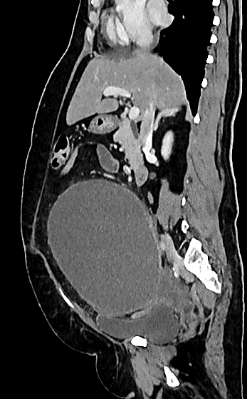
Figure 1: Contrast enhanced CT sagittal view - a large cyst, extending from the pelvis to the epigastrium without septations or solid components, possibly ovarian in origin.
Treatment
After optimization of her medical comorbidities, the patient underwent laparotomy. Three liters of foul smelling, muddy fluid was aspirated from a cyst, 20x10cm in size, to which the small bowel and omentum were adherent (Figure 2). The uterus was absent, the patient having undergone hysterectomy for fibroids 15 years earlier. Both ovaries were found adjacent to the cyst and were removed with the fallopian tubes. Portions of the rectus sheet was thickened and calcified, almost bone like (Figure 3). The omentum felt fibrous and the infracolic portion was excised as well.
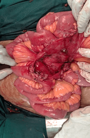
Figure 2: Intraoperative image - paraovarian cyst adherent to bowl.
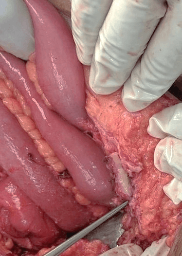
Figure 3: Intraoperative image - bony transformation of rectus sheath.
Outcome
The patient required careful monitoring and inotropic support in an intensive care ward in the postoperative period. Digoxin was added once weaned off inotropes. The fluid aspirated from the cyst grew Salmonella group B and ceftriaxone was continued in the postoperative period for an additional 14 days, as per antimicrobial susceptibility patterns. The patient continued to have occasional loose stools although microscopic examination of the stool did not reveal any anaerobes, ova or cysts and stool cultures did not grow Salmonella or any other enteric pathogen. Histological examination revealed that cyst was paraovarian, a simple serous cystadenoma. A portion of rectus sheath excised was found to be bony tissue. The removed omentum showed extensive areas of fibrosis with foamy histiocytes with few scattered blood vessels-features of sclerosing peritonitis.
Wound dehiscence of the muscle and rectus layers was discovered ten days after surgery, possibly a consequence of poor healing of the osseous metaplasia of the rectus sheet. The wound underwent secondary re-suturing and the patient was discharged. She remained symptom free on follow up one month later.
Discussion
Reports on suppuration of ovarian cysts by typhoid, gonococcal and mycobacterial bacillus are few but date back to the late 1800s. Bland-Sutton wondered “how ovarian cysts should become inflamed, enclosed as they are in air-tight cavities, and having no communication with other organ”. Pfannenstiel opined that infection must originate from the fallopian tubes or intestines – especially, in cases where the latter was adherent to the cyst. Taylor et al. in 1907 published a series of reports of typhoid infection of ovarian cysts [4]. In many cases, the febrile illness had occurred several weeks prior to the presentation with pain and mass per abdomen. The author concluded that since the bacilli rapidly disappear from the blood stream, infection of these cysts must have occurred during the acute bacteremic stage of the disease, and the bacilli must have been dormant in their local nidus since the attack, which may have occurred months or even years before. In all cases, the cysts were to be adherent to the bowel or omentum, suggesting that this is the route of transmission.
There have been several reports of infection of ovarian cysts by Salmonella [5-12]. Focal metastatic Salmonella infections are seen in almost every site in the body, attracted by a pre-existent ovarian lesion, or simply by the immunologically privileged ovary. Reports of infected ovarian abscesses in adolescents without sexual contact, suggest a hematogenous or gastrointestinal mode of spread, as hypothesized by Taylor [5, 8, 9]. This route of infection is also likely in our patient, where a paraovarian cyst was infected and the adjacent fallopian tubes did not show evidence of inflammation. A review of 33 cases of Salmonella ovarian abscesses revealed that in 79%, blood cultures were sterile, as in our case [6].
Another interesting feature in this case was the sclerosing peritonitis and osseous metaplasia of the rectus sheet, possibly another response to a disseminated infection of non-typhoidal Salmonella. Sclerosing peritonitis is a fibrous thickening of the parietal and visceral peritoneum, first described in 1907 and sometimes referred to as “abdominal cocoon” [13]. Sclerosing peritonitis maybe primary, a developmental anomaly. This is an accessory peritoneal membrane, derived from the yolk sac peritoneum, a predominately asymptomatic condition and is generally detected incidentally during a laparotomy performed for some other purpose. Secondary sclerosing peritonitis is a consequence of peritoneal inflammation due to various triggering factors: drug related, following intra-abdominal procedures such as peritoneal dialysis or infections a such as abdominal tuberculosis. The calcified rectus sheath in our patient which was histologically identified as bone, could be a consequence of the peritoneal inflammation. Compounded by her diabetes, the ossified rectus healed poorly, leading to burst abdomen in the post-operative period.
Hence, our patient presented with rare complication of an emerging disease. A depressed immune status, influenced by her comorbidities, and the lack of sanitation in pockets of South Asia may have led to a reappearance of these once historic complications of typhoid.
Conflicts of Interest
None.
Funding
None.
Article Info
Article Type
Case ReportPublication history
Received: Fri 17, Jul 2020Accepted: Wed 29, Jul 2020
Published: Mon 10, Aug 2020
Copyright
© 2023 Saroj Rajan. This is an open-access article distributed under the terms of the Creative Commons Attribution License, which permits unrestricted use, distribution, and reproduction in any medium, provided the original author and source are credited. Hosting by Science Repository.DOI: 10.31487/j.SCR.2020.08.09
Figures & Tables
References
- Stanaway JD, Reiner RC, Blacker BF, Goldberg EM, Khalil IA et al. (2019) The global burden of typhoid and paratyphoid fevers: a systematic analysis for the Global Burden of Disease Study 2017. Lancet Infect Dis 19: 369-381. [Crossref]
- Stanaway JD, Parisi A, Sarkar K, Blacker BF, Reiner RC et al. (2019) The global burden of non-typhoidal salmonella invasive disease: a systematic analysis for the Global Burden of Disease Study 2017. Lancet Infect Dis 19: 1312-1324. [Crossref]
- Patra S, Mukim Y, Varma M, Mukhopadhyay C, Kalwaje Eshwara V (2018) Invasive nontyphoidal Salmonella disease in southern India: a 5-year experience from a tertiary care hospital. Turk J Med Sci 48: 1030-1035. [Crossref]
- Taylor FE (1907) Typhoid Infection of Ovarian Cysts*. BJOG: Int J Obstet Gynaecol 12: 367-383.
- Getahun SA, Limaono J, Ligaitukana R, Cabenatabua O, Soqo V et al. (2019) Ovarian abscess caused by Salmonella enterica serovar Typhi: a case report. J Med Case Rep 13: 303. [Crossref]
- Adelman MW, Johnson JH, Hohmann EL, Gandhi RT (2017) Ovarian Endometrioma Superinfected With Salmonella: Case Report and Review of the Literature. Open Forum Infect Dis 4: ofx048. [Crossref]
- Kudesia R, Gupta D (2011) Pelvic Salmonella Infection Masquerading as Gynecologic Malignancy. Obstet Gynecol 118: 475-477. [Crossref]
- Legay L, Ghiducci MC, Camus E (2019) Salmonella enteridis ovarian abscess in a 10-year-old patient in New Caledonia - Case report and review of the literature. J Pediatric Surg Case Rep 49: 101275.
- Gorisek NM, Oreskovic S, But I (2011) Salmonella ovarian abscess in young girl presented as acute abdomen--case report. Coll Antropol 35: 223-225. [Crossref]
- Sharma P, Bhuju A, Tuladhar R, Parry CM, Basnyat B (2017) Tubo-ovarian abscess infected by Salmonella typhi. BMJ Case Rep 2017: bcr2017221213. [Crossref]
- Tohya T, Yoshimura T, Onoda C (2003) Unilateral Ovarian Abscess Caused by Salmonella. Infect Dis Obstet Gynecol 11: 217-219. [Crossref]
- Bjornson G, Scheifele DW, Bettinger J, Patrick DM, Gustafson L et al. (2007) Effectiveness of pneumococcal conjugate vaccine in Greater Vancouver, Canada: 2004-2005. Pediatr Infect Dis J 26: 540-542. [Crossref]
- Machado NO (2016) Sclerosing Encapsulating Peritonitis: Review. Sultan Qaboos Univ Med J 16: e142-e151. [Crossref]