
Journals
Neoplastic Seeding in Abdominal Wall Following Radiofrequency Ablation of Colorectal Liver Metastasis: Case Report
A B S T R A C T
Colorectal liver metastasis (CRLM) is the most common lesion diagnosed in the liver worldwide and new therapies have been proposed over the last decades. On the leading edge of these therapies, radiofrequency ablation (RFA) has been increasingly applied for treatment these lesions. The procedure can be done by percutaneous approach guided by CT, or during open or laparoscopic surgery. Although findings in preliminary reports suggest that this procedure is both safe and effective, only a few studies have been published. We present a 69-year-old female, previously submitted to two hepatic resections and one RFA for treatment of CRLM, who developed an abdominal wall recurrence due to neoplastic seeding after RFA procedure. In the published literature thus far, some risk factors have been associated with neoplastic seeding following RFA, like subcapsular location, multiple electrode placements, multiple RFA treatments and poor tumor differentiation grade. Technical features like access through normal liver substance and tract needle ablation may play a role in mitigating the risk of this complication. Although it seems to be a safe procedure, only a few retrospective papers have been published its rare complications and outcomes. Nevertheless, patients with localized disease may benefit from surgical resection.
Keywords
Colorectal liver metastasis, radiofrequency ablation, neoplastic seeding
Introduction
Colorectal cancer is a public health problem with a growing incidence worldwide. Fifty percent of patients will present a liver metastasis at some time during follow up. Although liver resection is currently considered the gold standard treatment for these lesions, minimally invasive therapy modalities have been proposed in many scenarios, whether for palliative or curative treatment. On the leading edge of these therapies, radiofrequency ablation (RFA) has been increasingly applied for treatment of colorectal liver metastasis (CRLM). The procedure can be done by percutaneous approach guided by CT, or during open or laparoscopic surgery. Although findings in preliminary reports suggest that this procedure is both safe and effective, only a few studies, have been published. Complications related to RFA include hepatic abscess, uncontrolled bleeding, intestinal perforation, biliary stenosis, hemobilia, and neoplastic seeding along puncture pathway.
The aim of this paper is to report a case of an abdominal wall resection after neoplastic seeding following RFA of CRLM and review the literature regarding RFA for resectable and unresectable disease.
Case Report
A 69-year-old female with a past history of low rectal cancer diagnosed in February 2012, treated initially with exclusive chemoradiotherapy with pathological response. After 48 months during the follow up, a rectal mass with synchronic liver metastasis were arised. After that, she was submitted to abdominoperineal resection of the primary tumor, followed by adjuvant chemoterapy and right hepatectomy 6 months later. In September 2017, with the CEA’s value increasing, a new restaging was done and a tumor recurrence of 2.6cm diameter in liver-segment III was noted, which was then treated by percutaneous RFA and this procedure was performed using internally-cooled electrodes (Cool-tip RF system; Valleylab, Boulder, CO, USA) with sedation and CT-guided.
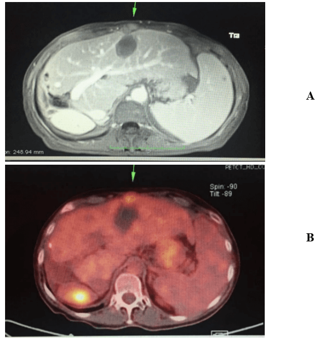
Figure 1: A) CT scan showing the necrotic nodular area in liver-segment III and the hipervascular lesion in the abdominal wall above it (green arrow). B) 18FDG-PET/CT showing the hypercaptation (green arrow).
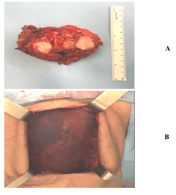
Figure 2: A) Surgical specimen showing de parietal nodule and its relationship with the liver surface. B) Abdominal wall reconstructed with a 10cmx20cm substitution mesh (Sepramesh®).
After 11 months, patient developed a tumoral lesion in the abdominal wall, immediately above de necrotic area of the RFA, suggesting neoplastic seeding in the puncture pathway (Figure 1A). A whole-body 18FDG-PET/CT confirmed hypercaptation only in the parietal nodule (Figure 1B). It was performed an abdominal wall resection, including the skin, subcutaneous fat tissue, abdominal rectus muscle and peritoneum with a carcinoma negative margins in frozen section specimen (Figure 2A). A 10cmx20cm substitution mesh (Sepramesh®) was used for wall reconstruction (Figure 2B), the patient had an uneventful postoperative course and was discharged on the 3rd postoperative day. Patient remains in follow up with the multidisciplinary team, with good performance status.
Discussion
Many papers have been published regarding the efficacy and results of RFA for CLRM, and this procedure has been rapidly adopted in the last decades in favor of other minimally invasive therapies, such as percutaneous ethanol injection (PEI), and in some cases, it has been chosen over surgery because of its potential benefits, including reduced morbidity and mortality. Indeed, it can be performed multiple times because is a parenchyma-sparing procedure. However, there are still few reports from small retrospective studies regarding the prevalence of its complications. Additionally, the complication rate of RFA must be compared with the morbidity and mortality rates of surgery (15-30% and 5%, respectively), as well as to the complication rate for PEI (with a mortality up to 0.7% and a major complication rate of 3.2%-4.6%) to permit an objective assessment of the risks versus benefits of this technology.
Neoplastic seeding of the needle path has been a known complication since the introduction of percutaneous tumor biopsy and therapy. At large, the rate of needle path during RFA of hepatic malignancy is up to 12.5% but for CRLM is less well defined [1].
Kosari et al., analyzing patterns of recurrence in 143 patients submitted to RFA for hepatic malignancies, reported a local recurrence rate of 7.7 %, compared to 51 % systemic (hepatic and extrahepatic) recurrence, with no needle path recurrence [2]. Patients with colorectal metastatic lesions > 4 cm submitted to RFA were more likely to present with local recurrence, and size was the only variable that was statistically significantly associated with this result. Although no needle path recurrence has been related, and no comparison could be done, the ablation of needle path by the probe tip during removal may play a role on preventing this complication. Besides that, all lesions were accessed through normal liver substance to avoid the risk of needle-related free peritoneal tumor seeding. From a practical qualitative standpoint, the more difficult lesions to treat were sclerotic in character (renal cell and colorectal metastases specifically) [2].
There are some risk factors associated with neoplastic seeding following RFA for treatment to Hepatocellular carcinoma (HCC), like subcapsular location, multiple electrode placements, multiple RFA treatments, poor tumor differentiation grade that we do not find out described for RFA to CRLM, in the literature [1, 3]. In both types of RFA indication tumors, HCC and CRLM, the subcapsular location is an important risk factor for seeding. Jaskolka et al., found an Odds Ratio=11.57 with p=0.007 in their series and Llovet et al. noted the same fact with statistically significant date, and proposed do not indicate RFA for subcapsular tumors and showed one more distinct point yet, the high risk for RFA with cooled-tip needle, the same used in this case report [1, 3]. There are three papers in the literature review regarding skin seeding after RFA for CRLM [4-6]. Bonatti et al. and Charalampopoulos et al. tried this complication after the second RFA session, showing us what Jaskolka et al. described, in 2005, with the HCC treatment [3-5]. All of four case reports, including our case, the recurrence was less than 12 months.
To prevent or decrease the neoplastic seeding risk, in the end of procedure it can be done the track ablation during the RFA needle’s withdrawal. For recurrence treatment, the resection with wide margin may be the best choice to keep the patient without disease.
Conclusion
Radiofrequency ablation is a relatively recent treatment in multimodality approach of CRLM. It plays a special hole in specific scenario, as in patients unfit for surgery, patients submitted previously to multiple abdominals procedures or in addition to multiple hepatic resection, in order to preserve parenchyma. Although it seems to be a safe procedure, only a few retrospective papers have been published its rare complications and outcomes. Regarding neoplastic seeding along puncture pathway, there are few reports thus far in the literature, but patients with localized disease may benefit from surgical resection.
Article Info
Article Type
Case ReportPublication history
Received: Fri 10, Jul 2020Accepted: Tue 21, Jul 2020
Published: Mon 03, Aug 2020
Copyright
© 2023 Reinaldo Fernandes. This is an open-access article distributed under the terms of the Creative Commons Attribution License, which permits unrestricted use, distribution, and reproduction in any medium, provided the original author and source are credited. Hosting by Science Repository.DOI: 10.31487/j.SCR.2020.08.03
Author Info
Corresponding Author
Reinaldo FernandesSurgeon, Hepatobiliary Multidisciplinary Team, Brazil
Figures & Tables
References
- J M Llovet, R Vilana, C Brú, L Bianchi, J M Salmeron et al. (2001) Increased risk of tumor seeding after percutaneous radiofrequency ablation for single hepatocellular carcinoma. Hepatology 33: 1124-1129. [Crossref]
- Kambiz Kosari, Maria Gomes, David Hunter, Donavan J Hess, Edward Greeno et al. (2002) Local, Intrahepatic, and Systemic Recurrence Patterns After Radiofrequency Ablation of Hepatic Malignancies. J Gastrointest Surg 6: 255-263. [Crossref]
- Jeffrey D Jaskolka, Murray R Asch, John R Kachura, Chia S Ho, Marc Ossip et al. (2005) Needle tract seeding after radiofrequency ablation of hepatic tumors. J Vasc Interv Radiol 16: 485-491. [Crossref]
- H Bonatti, G Bodner, P Obrist, O Bechter, G Wetscher et al. (2003) Skin implant metastasis after percutaneous radio-frequency therapy of liver metastasis of a colorectal carcinoma. Am Surg 69: 763-765. [Crossref]
- A Charalampopoulos, A Macheras, E Misiakos, A Batistatou, D Peschos et al. (2007) Thoracoabdominal wall tumour seeding after percutaneous radiofrequency ablation for recurrent colorectal liver metastatic lesion: a case report with a brief literature review. Acta Gastroenterol Belg 70: 239-242. [Crossref]
- Shirley Yuk Wah Liu, Kit Fai Lee, Paul Bo San Lai (2009) Needle track seeding: a real hazard after percutaneous radiofrequency ablation for colorectal liver metastasis. World J Gastroenterol 15: 1653-1655. [Crossref]