Longitudinal Melanonychia Treatment with Picosecond Laser for Differential Diagnosis
Longitudinal Melanonychia Treatment with Picosecond Laser for Differential Diagnosis
A B S T R A C T
Longitudinal melanonychia can have multiple causes, among them subungual melanoma. Clearing the pigmentation with pigment specific lasers, especially picosecond laser with less pain can be a comfortable and reliable alternative to ablation in the differential diagnosis.
Keywords
Melanonychia, nail, melanoma, pico laser
Since 2 years a brown linear hyperpigmentation on the left thumb nail without involvement of the nail edges (negative Hutchinson sign) persisted in a female Caucasian patient, age 49 years (Figure 1). No symptoms of internal and genetic diseases or dermatoses of the nail bed where documented. The negative Hutchinson sign did not support the suspicion of malignant melanoma. Linear nail pigmentations of this type are called longitudinal melanonychia [1]. The activity behind this symptom is caused by melanocytes on the distal end of the nail plate, that transfer melanin to onychocytes and by growing proximally, a linear pigmentation appears [2, 3].
Melanonychia is mostly benign. It can be initiated due to any activation of the nail matrix following for instance, melanin-producing pathogens, inflammation, or irritation of the nail and can be associated with melanocytic hyperplasia. Most patients with darker skin types show this symptom on the nails increasing with age equally in both sexes (100 % > 50 years, 77 % at > 20 years).
Common causes of longitudinal melanonychia include subungual hematomas and verrucae, nail trauma, onychomycosis nail psoriasis, lichen planus, chronic paronychia, pyogenic granuloma, Addison's disease, genetic causes like familial amyloidosis, Laugier-Hunziker syndrome, Peutz-Jeghers syndrome, Touraine syndrome [3-5]. But also, neoplasms, glomus tumor, keratoacanthoma, myxoid cysts and subungual melanoma.
Physical examination is difficult and includes: Variations in the colour and thickness, involvement of more than two-thirds of the nail plate, blurred borders larger than 3 millimeters, distortion of the nail plate, recurrent, spontaneous bleeding at the same site. The malignancy mainly affects people over 50 and is considered rare, accounting for only 0.7% to 0.35% of all skin cancers [3, 5, 6]. Subungual melanoma almost always appears on a single-digit only. One of the main clinical signs is the "Hutchinson's sign" a linear pigmentation running from nail bed to the distal end of the nail.
Unfortunately, patients with melanonychia are often initially misdiagnosed and nail melanoma carries a poor prognosis [4]. The 5-year and 10-year survival rates is reported to be 30% and 13%, respectively [5, 6]. For differential diagnosis the ablation of parts or the entire nail is necessary. Instead of a biopsy or the ablation, the laser reduction of the pigmentation helps in clinical differential diagnosis without damaging the nail and avoids cosmetical damage. So, we performed laser therapy in one single session with a picosecond laser (Pico Care, Won Tech, Korea) at 350 ps pulse duration, 532 nm, fluence 0, 5 J, spot size: 3 mm, 2 Hz and a total of 72 shots.
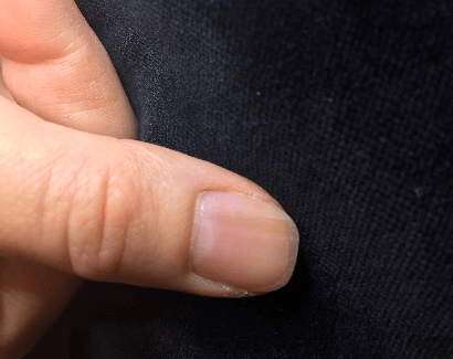
Figure 1: Longitudinal melanonychia on the left thumb in a 49-year-old Caucasian female patient before treatment.
The treatment was very well tolerated and resulted in a 95% pigment reduction after this one single session without any damage of the nail and without side effects and no reoccurrence until a follow-up of six months post treatment. Clearing pigmentation with pigment specific lasers, especially picosecond laser, which does not heat the nail as much as nanosecond lasers and because of this does not cause pain, are comfortable and reliable alternative to ablation and differential diagnosis.
Conflicts of Interest
None.
Article Info
Article Type
Case ReportPublication history
Received: Thu 20, Aug 2020Accepted: Tue 08, Sep 2020
Published: Fri 18, Sep 2020
Copyright
© 2023 Klaus Fritz. This is an open-access article distributed under the terms of the Creative Commons Attribution License, which permits unrestricted use, distribution, and reproduction in any medium, provided the original author and source are credited. Hosting by Science Repository.DOI: 10.31487/j.SCR.2020.09.09
Figures & Tables
References
- M G Haufs, O M Mainusch, J M Raguz, E Haneke (2001) Longitudinal melanonychia. Diagnosis, differential diagnosis and therapy. Dtsch Med Wochenschr 126: 561-564. [Crossref]
- Uwe Wollina, Pietro Nenoff, Gunter Haroske, Holger A Haenssle (2016) The Diagnosis and Treatment of Nail Disorders. Dtsch Arztebl Int 113: 509-518. [Crossref]
- Julie Jefferson, Phoebe Rich (2012) Melanonychia. Dermatol Res Pract 2012: 952186. [Crossref]
- Josette André, Nadine Lateur (2006) Pigmented nail disorders. Dermatol Clin 24: 329-339. [Crossref]
- Rich P (2006) Nail surgery. In: Bolognia JL, Jorizzo JL, Rapini RP, editors. Dermatology. 2nd edition. chapter 149. New York, NY, USA: Mosby. 2260-2268.
- Nathaniel Jellinek (2007) Nail matrix biopsy of longitudinal melanonychia: diagnostic algorithm including the matrix shave biopsy. J Am Acad Dermatol 56: 803-810. [Crossref]