
Journals
Long-term survival of patients with ischemic cardiomyopathy and diabetes as compared to those without diabetes undergoing myocardial viability assessment with 18FDG-PET
A B S T R A C T
Objectives: 18FDG-PET is considered the most sensitive test to detect hibernating myocardium. We compare the predictive value of 18FDG-PET for long-term survival of patients with ischemic cardiomyopathy and diabetes versus those without diabetes that were referred for 18FDG-PET to assess hibernating myocardium.
Patients and Methods: 80 patients (24 diabetics) with angiographically documented ischemic cardiomyopathy who underwent myocardial perfusion scintigraphy with 99mTc-tetrofosmin and myocardial viability evaluation by 18FDG PET/CT (after hyperinsulinemic-euglycemic clamp protocol with acipimox) were investigated. Median follow-up after viability testing was 62.4±33.2 months.
Results:All patients had impaired left ventricular ejection fraction (mean 32.9±9.3%). Diabetic patients had fewer percent of scar tissue (16.0±12.0) as compared to nondiabetic patients (25.7±18.7) (p < 0.01), while the amount of hypoperfused and viable myocardium was not significantly different (14.5±12.6 vs. 9.8±12.0) (p=0.21).12 patients had 1 vessel disease (VD), 15 patients 2 VD, 49 patients 3 VD, and 4 patients diffuse calcification in stenotic coronary arteries. Diabetic patients had significantly more commonly 3 VD than nondiabetic patients (83% vs. 52%, p < 0.01). The number of revascularization procedures in diabetic patients and nondiabetic patients was statistically not significantly different (71% vs. 54%, p=0.55). Median survival was 69.3 months (60.1-77.9 months) in nondiabetic patients and 46.5 months (32.7-60.3 months) in diabetic patients (p<0.001).
Conclusions: Patients with ischemic cardiomyopathy, impaired LVEF and diabetes exhibited significantly reduced overall survival compared to those without diabetes though having less scared and equal amounts of viable myocardium as indicated by 18FDG-PET.
K E Y W O R D S
Coronary disease, Diabetes, 18FDG-PET, Survival.
Introduction
Coronary artery disease (CAD) complicated by severe impaired left ventricular function is associated with high morbidity and mortality. Coronary revascularization may lead to symptomatic and prognostic improvement in such patients [1,2]. However, prospective identification of patients with ischemic cardiomyopathy and heart failure who may benefit from high risk revascularization remains a clinical challenge [1-3]. Hibernating myocardium, i.e., chronically hypoperfused myocardium leading to a hypocontractile state, is likely to benefit from revascularization as opposed to scarred myocardium. Reperfusion is known to potentially reverse wall motion abnormalities and improve left ventricular function of viable myocardium [1, 3]. Therefore, myocardial viability assessment is increasingly being used to identify CAD patients who are most likely to benefit from coronary revascularization procedures.
To detect the presence of viable myocardium, stress echocardiography, single-photon-emission computed tomography (SPECT) [1, 3] and more recently 18F-fluordesoxyglucose (FDG)-positron emission tomography (PET) have been used. Recently data from a study in 601 patients did not confirm the prognostic value of viability assessment by SPECT or stress echocardiography for survival or efficacy of coronary revascularization [1]. 18FDG-PET has been shown to have the highest sensitivity to detect hibernating myocardium [3,4], and CAD patients were found to benefit from PET-assisted management [5, 6]. 18FDG-PET seems to define high-risk patients that gain benefit from coronary revascularization. However, little agreement exists about the amount of viable myocardium needed to achieve a relevant improvement of left ventricular ejection fraction (LVEF) of > 5 %. Proposed cut-off-points range from 7% to 37% of left ventricular area [7].
Diabetic patients have increased mortality and morbidity from cardiovascular diseases, independent of other risk factors [8-10]. Revascularization procedures are associated with a higher morbidity and mortality in patients with diabetes mellitus than in nondiabetic patients [11].
In this study, we investigate retrospectively the long-term survival of patients with ischemic cardiomyopathy, impaired LVEF and diabetes as compared to those without diabetes that were referred for myocardial viability assessment with 18FDG-PET.
Methods
A cohort of eighty consecutive patients (71 males and 9 females, mean age of 71.3±9.8 years) with predominantly symptoms of heart failure that had angiographically documented ischemic cardiomyopathy, fixed perfusion defects in 99mTc-tetrofosmin SPECT, impaired LVEF and were referred for 18FDG-PET for viability assessment to detect hibernating myocardium at our Institution between 2011 and 2015 were included in this study. Patients with deaths attributed to non-cardiac causes were excluded from this investigation. Fifty-four patients had previous acute myocardial infarction.
All patients underwent myocardial perfusion scintigraphy using 99mTc-tetrofosmin and evaluation of myocardial viability by 18FDG-PET/CT using a combination of hyperinsulinemic-euglycemic clamp protocol with acipimox. Inclusion criteria for 18FDG-PET imaging were wall motion abnormalities in previous clinical echocardiographic evaluation, fixed perfusion defects in 99mTc-tetrofosmin SPECT and LVEF < 50%. Median time of follow-up (performed with conventional protocols for cardiac patients) was 62.4±33.2 months after viability testing. All patients signed an informed consent. The study was in accordance with the declaration of Helsinki and approved by an institutional review board.
Myocardial perfusion was assessed with 99mTc-tetrofosmin (4 MBq/kg body weight) gated SPECT using a two-day stress/rest protocol and pharmacologic stress with adenosine (140 μg/kg body weight/minute for 4 minutes). Acquisition (rotation 90°, 34 s/projection, 64x64 matrix) was done using a Siemens E.CAM gamma camera with a low-energy high-resolution-collimator. Quantitative myocardial perfusion analysis was performed on a 17-segment model using 4 DM-SPECT software.
Metabolic activity was measured with 18FDG-PET (4.2 MBq/kg body weight), following a euglycemic-hyperinsulinemic protocol (250 mg acipimox at least one hour before tracer injection). A Siemens Biograph 6 PET-Scanner, 168x168 matrix, iterative reconstruction and 5 mm FWHM filter were used. Myocardial viability assessment was based on the concept of perfusion-metabolism mismatch found in myocardium with previously determined contractile dysfunction. Mismatch was expressed as a percentage of the left ventricle.
Revascularization was intended in our Institution when the amount of hypoperfused but viable myocardium exceeded 10% of left ventricular area. However, we included also in this study 13 patients with coronary artery stenosis and myocardial viability < 10% that have been revascularized at other institutions, and 3 other patients with myocardial viability >20% that have refused revascularization in our Institution.
Statistical analysis
Data consistencies were checked, and data were screened for outliers and normality using quantitative plots. Potential confounders were adjusted for survival analysis. Two-sided, independent t-tests with and without the assumption of variance homogeneity were used to compare various variables between both groups. The association between overall survival and diabetes was assessed by applying Cox proportional models and by computing Cox-Mantel hazard ratios together with 95% CI. The assumption of proportional hazard ratios was tested by corresponding tests. The hazard ratios were tested using the Cox-Mantel Log rank test. All reported tests were two-sided, and p-values < 0.05 were considered as statistically significant. All statistical analyses in this report were performed by use of NCSS (NCSS 10, NCSS, LLC, Kaysville, UT) and STATISTICA 12 (Hill, T & Lewicki).
Results
Clinical data of patients are shown in Table 1. No significant differences between diabetic and nondiabetic patients were found in clinical data (i.e. age, cigarette smoking, arterial hypertension, hyperlipidemia, renal insufficiency and LVEF) and treatment (patients that underwent revascularization and patients who received medical therapy alone).
Table 1: Clinical data of patients
|
|
Patients with diabetes (n=24) |
Patients without diabetes (n=56) |
Patients with revascularization (n=47) |
Patients with medical therapy alone (n=33) |
|
Age (years) (range, mean) |
56 – 80 (68.6) |
50 – 82 (70.3) |
50 – 82 (68.3) |
56 – 81 (70.7) |
|
Gender |
20 male, 4 female |
51 male, 5 female |
43 male, 4 female |
28 male, 5 female |
|
Cigarette smoking (n=27) |
9 |
18 |
19 |
8 |
|
Arterial hypertension (n=51) |
15 |
36 |
32 |
19 |
|
Hyperlipidemia (n=61) |
18 |
43 |
39 |
22 |
|
Renal insufficiency (n=16) |
5 |
11 |
9 |
7 |
|
LVEF (%) (range, mean) |
19 – 48 (35.8) |
15 – 48 (31.6) |
15 – 48 (33.2) |
17 – 48 (31.2) |
n, number of patients; LVEF, left ventricular ejection fraction
All patients had impaired LVEF (mean 32.9±9.3, range 15-48%). Diabetic patients had fewer percent of scar tissue (16.0±12.0) as compared to nondiabetic patients (25.7±18.7) (p < 0.01), while the amount of hypoperfused but viable myocardium was not significantly different (14.5±12.6 vs. 9.8±12.0) (p=0.21) (Figures 1 and 2). No patient showed reverse perfusion-metabolism mismatch. In 3 out of 24 patients with diabetes, 18FDG-myocardial uptake was diffusely reduced which although resulted in poor-quality scans still enabled interpretation of the images and therefore further inclusion of these patients in this study.
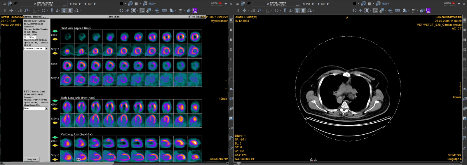
Figure 1: Small non-transmural scar in the anteroseptal distal wall and viable myocardium in all other segments in a 68 years old male patient with diabetes and LVEF of 27 % who died 52.8 months after myocardial viability assessment.
GRst, gated rest; Stdy-2, myocardial FDG PET.
Twelve patients had angiographically documented 1 vessel disease (VD), 15 patients 2 VD, 49 patients 3 VD, and 4 patients diffuse calcification in stenotic coronary arteries. Patients with diabetes had significantly more commonly 3 VD than patients without diabetes (83% vs. 52%, p < 0.01). Forty-seven patients underwent coronary revascularization [coronary-artery bypass grafting (CABG), 24 patients; stenting, 23 patients] and 33 patients’ medical therapy alone. Twenty-four out of 80 patients had diabetes mellitus type II. The number of coronary revascularization procedures in patients with and without diabetes was statistically not significantly different (71% vs. 54%, p=0.55).
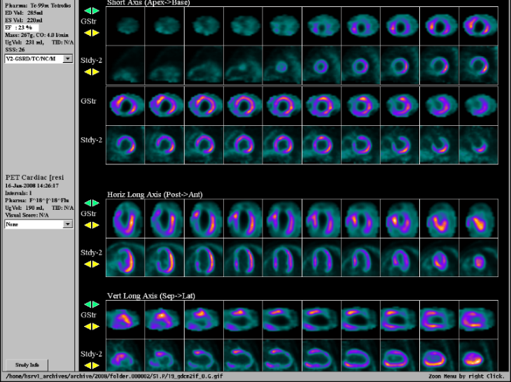
Figure 2: Large transmural scar in the apex and anterior and posterior distal wall and viable myocardium in all other segments in a 58 years old male patient without diabetes and LVEF of 29%, still alive 103.2 months after myocardial viability assessment. GSt, gated rest; Stdy-2, myocardial FDG PET.
Overall survival was 64% (51/80 patients). It was significantly favorable in patients without diabetes with a hazard ratio of 4.2 for these patients (95% CI: 1.77–9.9, p < 0.001) (Figure 3).
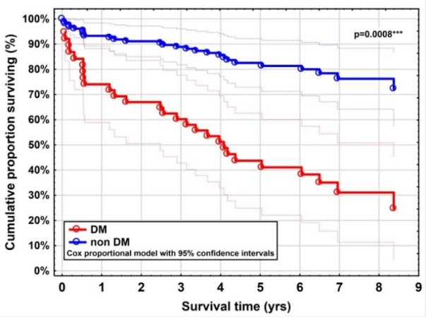
Figure 3: verall survival in patients with diabetes and patients without diabetes.
Median survival in nondiabetic patients (69.3 months, range 60.1-77.9 months) was significantly longer (p < 0.001) than that of patients with diabetes (46.5 months, range 32.7-60.3 months). No significant survival benefit for revascularization compared to medical therapy alone using viability cut-off-points of 10%, 20% and 30% was found both in diabetics (p=0.88) and nondiabetic patients (p=0.56). Longer survival was however noticed in patients with larger amounts of viable myocardium that underwent revascularization by CABG or stenting compared to patients who received medical therapy alone (Table 2).
Table 2: Results of survival according to myocardial viability cut-off-points
|
Survival |
Myocardial viability |
||||||
|
<10% |
>10% |
<20% |
>20% |
<30% |
>30% |
||
|
n=41 |
n=39 |
n=56 |
n=24 |
n=68 |
n=12 |
||
|
Overall survival |
30 (73%) |
29 (74%) |
42 (75%) |
17 (71%) |
48 (71%) |
11 (92%) |
|
|
Revascularized patients |
13 (76%) |
23 (77%) |
22 (79%) |
14 (74%) |
27 (71%) |
9 (100%) |
|
|
CABG |
5 (71%) |
14 (82%) |
10 (83%) |
9 (75%) |
14 (74%) |
5 (100%) |
|
|
Stenting |
8 (80%) |
9 (69%) |
12 (75%) |
5 (71%) |
13 (68%) |
4 (100%) |
|
|
Medically treated only patients |
17 (71%) |
6 (67%) |
20 (71%) |
3 (60%) |
21 (70%) |
2 (67%) |
|
Discussion
Despite improvement in the management of patients with CAD, the presence of diabetes mellitus has been found to remain associated with worse outcomes [8-10]. We observed that patients with diabetes had fewer percent of scar tissue than nondiabetic patients, while the amount of hypoperfused but viable myocardium was not significantly different. Noteworthy, we found a significant death rate in the first year and a significantly shorter overall survival in the diabetic group although the LVEF values in this group were not significantly different from those found in the nondiabetic group. These findings seem to indicate diabetes as a predictor for early mortality in patients with ischemic cardiomyopathy. Furthermore, in our patient population diabetic patients had significantly more commonly 3 VD than nondiabetic patients. The presence of 3 VD may have been associated with systemic vascular disease which might have contributed to the shorter survival found in our group of patients with diabetes.
Coronary revascularization has greatly improved the outcomes of CAD patients with diabetes [9, 11]. However, much work remains to better understand the underlying mechanisms of CAD in the setting of diabetes and to improve clinical outcomes in this patient population. The development of new treatment strategies and recognition of the association between diabetes and outcomes after revascularization may help identify novel treatments for this high-risk group of patients. Two meta-analyses showed that patients with viable myocardium who underwent coronary revascularization have longer survival than those receiving medical therapy alone [12, 13]. The PARR-2 trial reported that 18FDG-PET had a non-significant trend for improved outcome of coronary revascularization compared with standard care [14]. The STICH Extension Study (STICHES), which was conducted to evaluate the long-term effects of CABG in patients with ischemic cardiomyopathy found that the rates of death from cardiovascular causes, and death from any cause or hospitalization for cardiovascular causes were significantly lower over 10 years among patients who underwent CABG in addition to receiving medical therapy than among those who received medical therapy alone [15]. We found that none of the myocardial viability thresholds investigated showed significant prognostic value regarding survival for the benefit from revascularization compared to medical therapy alone. However, it is noteworthy in our study that, as the amount of myocardial viability increases, there is a progressive increase in survival when revascularization is undertaken compared with medical therapy alone. Interestingly, this trend was more evident in patients who underwent CABG that in patients receiving stenting. Limitations of this retrospective study are the small number of patients in our subgroups. Further randomized studies with a larger number of patients are ongoing to evaluate the correlation between the amount of ischemia revascularized and the outcome after revascularization in our patient population.
Data evaluating the relationship of viability extent and outcome response to coronary revascularization are limited. Survival was found shorter when mismatch was >20% and LVEF < 43% [16, 17]. D’Egidio et al. showed that patients with viability >7% gained an outcome benefit when revascularization was undertaken compared with medical therapy, while in patients with viability < 7% the outcome was not significantly different [7]. In our study, no significant differences were found in overall survival or survival in coronary revascularized patients as viability increased applying myocardial viability thresholds of 10%, 20% and 30%. However, in patients receiving medically treatment only, a non-significant trend for shorter survival when the extent of viability increased was observed. These findings may be attributed to the fact that our patient population had severely reduced LVEF (mean 32.9±9.3%).
An adequate regulation of metabolic conditions is needed to ensure optimal image quality with 18FDG-PET. However, insulin resistance is frequently present in patients with heart failure, and the amount of endogenous insulin released after oral glucose loading will not induce maximal stimulation of myocardial 18FDG uptake [18]. Our data confirm that myocardial imaging with 18FDG-PET following a euglycemic-hyperinsulinemic protocol with acipimox results in adequate imaging quality in the vast majority of diabetic patients.
Conclusions
Ischemic cardiomyopathy patients with impaired LVEF and diabetes exhibited significantly reduced overall survival compared to patients without diabetes though they had less scared myocardium and equal amounts of viable myocardium as indicated by 18FDG-PET.
No correlation was found between overall survival (both in diabetic and nondiabetic patients) and myocardial viability thresholds of 10%, 20% or 30%.
Conflicts of interest
There are no conflicts of interest.
Article Info
Article Type
Research ArticlePublication history
Received: Wed 26, Dec 2018Accepted: Tue 08, Jan 2019
Published: Sat 23, Feb 2019
Copyright
© 2023 Christian Pirich. This is an open-access article distributed under the terms of the Creative Commons Attribution License, which permits unrestricted use, distribution, and reproduction in any medium, provided the original author and source are credited. Hosting by Science Repository.DOI: 10.31487/j.JICOA.2019.01.001
Author Info
Corresponding Author
Christian PirichDepartment of Nuclear Medicine and Endocrinology, Paracelsus Medical University, Müllner Hauptstrasse 48, A-5020 Salzburg, Austria
Figures & Tables
GRst, gated rest; Stdy-2, myocardial FDG PET.
Table 1: Clinical data of patients
|
|
Patients with diabetes (n=24) |
Patients without diabetes (n=56) |
Patients with revascularization (n=47) |
Patients with medical therapy alone (n=33) |
|
Age (years) (range, mean) |
56 – 80 (68.6) |
50 – 82 (70.3) |
50 – 82 (68.3) |
56 – 81 (70.7) |
|
Gender |
20 male, 4 female |
51 male, 5 female |
43 male, 4 female |
28 male, 5 female |
|
Cigarette smoking (n=27) |
9 |
18 |
19 |
8 |
|
Arterial hypertension (n=51) |
15 |
36 |
32 |
19 |
|
Hyperlipidemia (n=61) |
18 |
43 |
39 |
22 |
|
Renal insufficiency (n=16) |
5 |
11 |
9 |
7 |
|
LVEF (%) (range, mean) |
19 – 48 (35.8) |
15 – 48 (31.6) |
15 – 48 (33.2) |
17 – 48 (31.2) |
n, number of patients; LVEF, left ventricular ejection fraction
Table 2: Results of survival according to myocardial viability cut-off-points
|
Survival |
Myocardial viability |
||||||
|
<10% |
>10% |
<20% |
>20% |
<30% |
>30% |
||
|
n=41 |
n=39 |
n=56 |
n=24 |
n=68 |
n=12 |
||
|
Overall survival |
30 (73%) |
29 (74%) |
42 (75%) |
17 (71%) |
48 (71%) |
11 (92%) |
|
|
Revascularized patients |
13 (76%) |
23 (77%) |
22 (79%) |
14 (74%) |
27 (71%) |
9 (100%) |
|
|
CABG |
5 (71%) |
14 (82%) |
10 (83%) |
9 (75%) |
14 (74%) |
5 (100%) |
|
|
Stenting |
8 (80%) |
9 (69%) |
12 (75%) |
5 (71%) |
13 (68%) |
4 (100%) |
|
|
Medically treated only patients |
17 (71%) |
6 (67%) |
20 (71%) |
3 (60%) |
21 (70%) |
2 (67%) |
|
References
1. Bonow RO, Maurer G, Lee KL, Holly TA, Binkley PF, et al. (2011) Myocardial viability and survival in ischemic left-ventricular dysfunction. N Engl J Med 364: 1617-1625. [Crossref]
2. Singh P, Sethi N, Kaur N, Kozman H (2015) Revascularization in severe left ventricular dysfunction: does myocardial viability even matter? Clin Med Insights Cardiol 9(Suppl 1): 105-109. [Crossref]
3. Schinkel AF, Bax JJ, Delgado V, Poldermans D, Rahimtoola SH (2010) Clinical relevance of hibernating myocardium in ischemic left-ventricular dysfunction. Am J Med 123: 978-986. [Crossref]
4. Arrighi JA (2006) Assessment of myocardial viability: more than measurements of radiotracer uptake alone. J Nucl Cardiol 13: 180-183. [Crossref]
5. Abraham A, Nichol G, Williams KA, Guo A, de Kemp RA, et al. (2010) F-18 FDG imaging of myocardial viability in an experienced center with access to F-18 FDG and integration with clinical management teams: the Ottawa-FIVE substudy of the PARR 2 Trail. J Nucl Med 51: 567-574. [Crossref]
6. Uebleis C, Hellweger S, Laubender RP, Becker A, Sohn HY, et al. (2013) The amount of disfunctional but viable myocardium predicts long-term survival in patients with ischemic cardiomyopathy and left ventricular dysfunction. Int J Cardiovasc Imaging 29: 1645-1653. [Crossref]
7. D Egidio G, Nichol G, Williams KA, Guo A, Garrard L, et al. (2009) Increasing benefit from revascularization is associated with increasing amounts of myocardial hibernation: a substudy of the PARR-2 trial. JACC Cardiovasc Imaging 2: 1060-1068. [Crossref]
8. Almdal T, Scharling H, Jensen JS, Vestergaard H (2004) The independent effect of type 2 diabetes mellitus on ischemic heart disease, stroke, and death: a population-based study of 13,000 men and women with 20 years of follow-up. Arch Intern Med 164: 1422-1426. [Crossref]
9. Armstrong EJ, Rutledge JC, Rogers JH (2013) Coronary artery revascularization in patients with diabetes mellitus. Circulation 128: 1675-1685. [Crossref]
10. Lima EG, Hueb W, Garcia RM, Pereira AC, Soares PR, et al. (2013) Impact of diabetes on 10-year outcomes of patients with multivessel coronary artery disease in the Medicine, Angioplasty, or Surgery Study II (MASS II) trial. Am Heart J 166: 250-257. [Crossref]
11. Herlitz J, Wognsen GB, Emanuelsson H, Haglid M, Karlson BW, et al. (1996) Mortality and morbidity in diabetic and nondiabetic patients during a 2-year period after coronary artery bypass grafting. Diabetes Care 19: 698-703. [Crossref]
12. Allman KC, Shaw L, Hachamovitch R, Udelson JE (2002) Myocardial viability testing and impact of revascularizationon prognosis in patients with coronary artery disease and left-ventricular dysfunction: a meta-analysis. J Am Coll Cardiol 39: 1151-1158. [Crossref]
13. Schinkel AF, Bax JJ, Poldermans D, Elhendy A, Ferrari R, et al. (2007) Hibernating myocardium: diagnosis and patient outcomes. Curr Probl Cardiol 32: 375-410. [Crossref]
14. Beanlands RS, Nichol G, Huszti E, Humen D, Racine N, et al. (2007) F-18-fluorodeoxyglucose positron emission tomography imaging-assisted management of patients with severe left-ventricular dysfunction and suspected coronary disease (PARR-2). J Am Coll Cardiol 50: 2002-2012. [Crossref]
15. Velazquez EJ, Lee KL, Jones RH, Al-Khalidi HR, Hill JA, et al. (2016) Coronary-artery bypass surgery in patients with ischemic cardiomyopathy. N Engl J Med 374: 1511-1520. [Crossref]
16. Desideri A, Cortigiani L, Christen A, Coscarelli S, Gregori D, et al. (2005) The extent of perfusion-F18-fluorodeoxyglucose positron emission tomography mismatch determines mortality in medically treated patients with chronic ischemic left-ventricular dysfunction. J Am Coll Cardiol 46: 1264-1269. [Crossref]
17. Yoshida K, Gould KL (1993) Quantitative relation of myocardial infarct size and myocardial viability by positron emission tomography to left-ventricular ejection fraction and 3-year mortality with and without revascularization. J Am Coll Cardiol 22: 984-997. [Crossref]
18. Schinkel AF, Bax JJ, Valkema R, Elhendy A, van Domburg RT, et al. (2003) Effect of diabetes mellitus on myocardial 18F-FDG SPECT using acipimox for the assessment of myocardial viability. J Nucl Med 44: 877-883. [Crossref]