
Infrahepatic Implantation of an Ectopic Pregnancy
A B S T R A C T
Background: Ectopic pregnancies occur predominantly in the fallopian tubes or ovaries. Very rarely, the distal part of the greater omentum may have close contact to the fallopian tubes and implantation of the embryo may occur in this part of the greater omentum. In the absence of signs of pregnancy in the uterus or the fallopian tubes, the greater omentum has to be closely examined for ectopic pregnancy.
Case Presentation: A 22-year-old woman in her 4th week of pregnancy presents with spotting and severe pain in the right lower abdomen with a history of 2 cesarean sections. The sonographic examination showed a normal uterus with a thin endometrial line. The uterine cavity did not present with any signs of a gestational sac. A great amount of free fluid in the Douglas cavity suspicious of a hemoperitoneum. ßhCG-values in serum was highly elevated up to 16749 mU/ml and confirmed the suspicion of an ectopic pregnancy. A diagnostic laparoscopy was performed, during which a blood clot reaching from the right lower abdomen to the greater omentum was detected. After removing the blood clot, a normal-sized uterus with regular ovaries and no signs of a pregnancy in the fallopian tubes were seen. However, an abdominal adhesion in the right upper part of the omentum close to the liver with bleeding was seen. While examining the abdominal cavity for the cause of bleeding, a small cystic lesion adherent to the greater omentum – close the offspring from the colon – representing an ectopic pregnancy became apparent. A partial omentectomy of 9,5 cm x 5,5 cm x 2,5 cm was performed with the finding of trophoblast and embryoblast implantated in the infrahepatic part of the greater omentum.
Conclusion: In cases of ectopic pregnancy with hemoperitoneum, special care has to be taken examining not only the fallopian tubes or ovaries but also the greater omentum, which may harbour an implanted trophoblast and embryoblast – even in the upper part directly beneath the liver – as presented in this case. In cases of ruptured ectopic pregnancy of the greater omentum, the cases may be dealt with laparoscopic partial omentectomy if the case is early detected.
Keywords
Ectopic pregnancy, infrahepatic implantation, hemoperitoneum
Introduction
Ectopic pregnancies occur in two percent of all pregnancies [1]. These rare ectopic implantations may cause diagnostic and therapeutic challenges [2]. Morbidity and mortality rates of ectopic pregnancies are still high, including massive blood loss, sepsis, systemic or organ failure, and death [3]. It needs to be considered that ectopic pregnancies not only may occur in the fallopian tubes but also in the cervix, the ovaries, the peritoneal cavity, and the omentum [2, 4]. These cases are difficult to detect and to remove, so it is critical to raise awareness of these unusual locations and also to confer strategies to cope with them.
Case Presentation
A 22-year-old woman in her 4th week of pregnancy presents with spotting and severe pain in the right lower abdomen. She had a history of 2 cesarean sections, the last one 4 months before the beginning of her symptoms, and referral to our outpatient clinic. The sonographic examination showed a normal uterus with a thin endometrial line. The uterine cavity did not present with any signs of a gestational sac. Next to the right ovary, a suspicious structure, potentially corresponding with an ectopic pregnancy of 30 mm x 33 mm, was detected. The left ovary had a normal size of 19 mm x 14 mm. A great amount of free fluid in the Douglas cavity was suspicious of a hemoperitoneum. ßhCG-values in serum were highly elevated up to 16749 mU/ml and confirmed the suspicion of an ectopic pregnancy. A diagnostic laparoscopy was performed, during which a blood clot reaching from the right lower abdomen to the greater omentum was detected. After removing the blood clot, a normal-sized uterus with regular ovaries and no signs of a pregnancy in the fallopian tubes became apparent (Figures 1 & 2). No signs of pregnancy in the fallopian tubes or ovaries were seen. Adhesions were identified between the uterus and the peritoneum of the bladder, and the anterior abdominal wall. However, an abdominal adhesion in the right upper part of the omentum close to the liver with bleeding was detected (Figure 3). While examining the abdominal cavity for the cause of bleeding, a small cystic lesion adherent to the greater omentum – close the offspring from the colon – representing a non-vital ectopic pregnancy was found. A partial omentectomy of 9,5 cm x 5,5 cm x 2,5 cm was performed (Figure 4). The histological examination revealed chorionic villi with a two-layer trophoblast with hyperemia and vasodilatation, confirming the diagnosis of ectopic pregnancy in the unusual infrahepatic location in the upper part of the greater omentum. During the following days after surgery, the ßhCG level decreased appropriately.
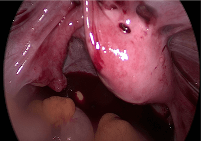
Figure 1: Ectopic pregnancy: normal left fallopian tube and adhesions of the uterus to the peritoneum of the bladder and the anterior abdominal wall originating from previous cesarean sections.
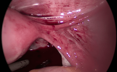
Figure 2: Ectopic pregnancy: normal right fallopian tube and hemoperitoneum (Douglas´ cavity).
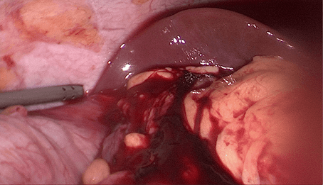
Figure 3: Suspicious hematoma in the upper part of the abdomen (infrahepatic).
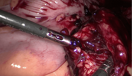
Figure 4: Laparoscopic partial omentectomy with the embedded ruptured ectopic pregnancy.
Discussion
Abdominal pregnancies defined by Studdiford in 1942 as a normal appearance of bilateral fallopian tubes and ovaries, the absence of uteroperitoneal fistula and a present pregnancy related to the peritoneal surface exclusively are very rare with only 1% of all ectopic pregnancies [5, 6]. The most common localisation of ectopic pregnancies is the fallopian tube. In only 5%, the implantation occurs in the ovaries, the peritoneum, the cervix, the pouch of Douglas, the pelvic sidewall, and the omentum [2, 4, 6-8]. Cases of abdominal pregnancy are described in literature mainly as case reports because of their rareness [9]. Besides artificial reproductive technologies, a history of ectopic pregnancy, pelvic inflammatory disease, current intrauterine pessaries, and tubal damage, the history of abdominal surgery in the present case with two cesarean sections seem to increase the risk for ectopic pregnancy [10, 11]. An implantation in an adhesion caused by prior surgeries can easily be dismissed. This increases the high morbidity and mortality rates of ectopic pregnancies [12]. Thus, precautions must be taken in cases of ectopic pregnancy, as they not only may occur in the fallopian tube or ovaries but also in the upper abdomen, especially in the greater omentum, which allows implantation and development of the embryo through it´s good blood supply. All efforts have to be taken to detect an ectopic pregnancy at the earliest stage possible. However, some abdominal pregnancy have been brought to full term. It is vital to closely examine all intraabdominal potential sites during diagnostic laparoscopy – not only fallopian tubes and ovaries but also the upper abdomen and the greater omentum as presented here – when abdominal pain and intra-abdominal bleeding occurs – and it is necessary to closely examine adhesions caused by prior surgeries. For better vascular control, the recommended treatment consists of exploratory laparotomy [9]. In this case, laparoscopy was the procedure performed since the source of bleeding was detected on time during the operation. In case of the suspicion of an ectopic pregnancy, a hemoperitoneum, and normal bilateral fallopian tubes and ovaries during exploration, surgeons should exclude an omental insertion of pregnancy. Adhesions of the uterus or the fallopian tubes after cesarean sections or other operations may interfere in the normal mechanism of uptake of the egg from the ovary and may favour the ectopic implantation of the embryo even to remote sites up to the greater omentum – infahepatic – as presented in this case.
Conclusion
Ectopic pregnancies and spontaneous abortions are difficult to detect. In cases of unsuspicious fallopian tubes and ovaries and no signs of implantation in the Douglas´ cavity, surgeons should always closely inspect the greater omentum for an unusual implantation in this site. With the risk of life-threatening blood loss, it is vital to raise the awareness of these unusual locations and also to confer strategies to manage them.
Conflicts of Interest
None.
Article Info
Article Type
Case ReportPublication history
Received: Sat 20, Feb 2021Accepted: Mon 15, Mar 2021
Published: Mon 03, May 2021
Copyright
© 2023 Peter Kern. This is an open-access article distributed under the terms of the Creative Commons Attribution License, which permits unrestricted use, distribution, and reproduction in any medium, provided the original author and source are credited. Hosting by Science Repository.DOI: 10.31487/j.SCR.2021.03.16
Figures & Tables
References
1.
Barnhart KT (2009) Clinical practice. Ectopic pregnancy. N
Engl J Med 361: 379-387. [Crossref]
2.
Stucki D, Buss J (2008) The ectopic pregnancy, a
diagnostic and therapeutic challenge. J Med Life 1: 40-48. [Crossref]
3.
Lisonkova S, Tan J, Wen Q, Abdellatif L, Richter LL et
al. (2019) Temporal trends in severe morbidity and mortality associated with
ectopic pregnancy requiring hospitalisation in Washington State, USA: a
population-based study. BMJ Open 9: e024353. [Crossref]
4.
Maiorana A, Incandela D, Giambanco L, Alio W, Alio L
(2014) Omental pregnancy: case report and review of literature. Pan Afr Med
J 19: 244. [Crossref]
5.
Studdiford WE (1942) Primary peritoneal pregnancy. Am
J Obstetr Gynecol 44: 487-491.
6.
Barel O, Suday RR, Stanleigh J, Pansky M (2019)
Laparoscopic Removal of an Abdominal Pregnancy in the Pelvic Sidewall. J
Minim Invasive Gynecol 26: 1007-1008. [Crossref]
7.
Thomas JB (2019) Rare primary abdominal ectopic pregnancy
in the pouch of Douglas. BMJ Case Rep 12: e228547. [Crossref]
8.
Memon Z, Sheikh SS (2015) Tubo-omental ectopic pregnancy.
J Pak Med Assoc 65: 215-217. [Crossref]
9.
Riethmuller D, Courtois L, Maillet R, Schaal JP (2003)
Ectopic pregnancy management: cervical and abdominal pregnancies. J Gynecol
Obstet Biol Reprod (Paris) 32: S101-S108. [Crossref]
10.
Yoder N, Tal R, Martin JR (2016) Abdominal ectopic
pregnancy after in vitro fertilization and single embryo transfer: a case
report and systematic review. Reprod Biol Endocrinol 14: 69. [Crossref]
11.
Thorburn J, Philipson M, Lindblom B (1986) Background
factors of ectopic pregnancy. II. Risk estimation by means of a logistic model.
Eur J Obstet Gynecol Reprod Biol 23: 333-340. [Crossref]
12. Srinivasan A, Millican S
(2014) Laparoscopic management of an abdominal pregnancy. Case Rep Obstet
Gynecol 2014: 562731. [Crossref]