Disseminated Pleural Malignant Melanoma
Case Presentation
A 68-year-old man was admitted to our hospital with a dyspnea. On physical examination the patient was not distressed. Chest X-ray demonstrated an extensive left pleural effusion (Figure 1A). Pleural aspiration showed an exsudate with elevated LDH of 464 U/L (0-100 U/l). The diagnostic thoracoscopy revealed disseminated black nodules on the visceral and parietal pleura (Figure 1B), and a pleurodesis was performed after obtaining multiple biopsies. The melanoma cells showed a strong cytoplasmatic HMB-45 reaction (Figure 1C).
Immunohistochemical staining revealed a malignant melanoma with expression of PD-L1 in 3% of the tumor cells, the molecular genetic examination proofed a BRAF V600 mutation. The patient was treated with the kinase inhibitor Trametinib and the tyrosinkinase inhibitor Tafiniar. He had initially a good response but died 10 months later after presenting to our clinic.
3 years prior the patient had a removal of a malign melanoma on the right upper thorax wall and the surgical exploration showed micro metastases in the sentinel lymph node of the right axilla. (SSM Clark Level IV). At that time, an immunotherapy with interferon alpha 2a was initiated.
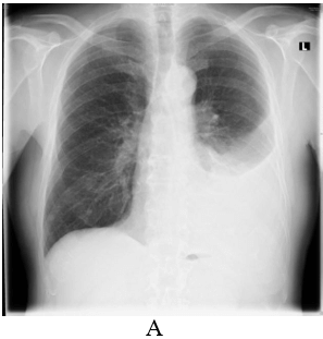
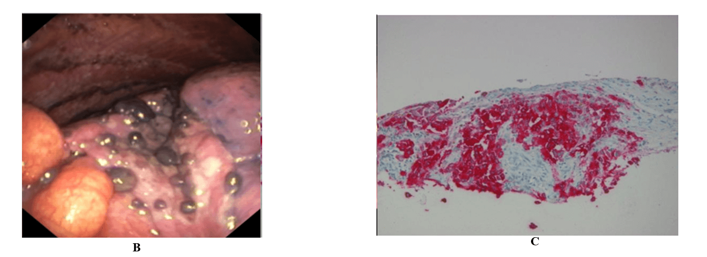
Figure 1: 4 years prior to his current hospital admission the patient had an excision of a malignant melanoma on the right shoulder.
A) Left pleural effusion. B) Disseminated black nodules on the visceral pleura. C) Immunohistochemical staining revealed a malignant melanoma with expression of PD-L1 in 3% of the tumor cells. The melanoma cells show a strong cytoplasmatic HMB-45 reaction.
Article Info
Article Type
Case ReportPublication history
Received: Mon 04, May 2020Accepted: Mon 15, Jun 2020
Published: Tue 30, Jun 2020
Copyright
© 2023 Jan Bronnert. This is an open-access article distributed under the terms of the Creative Commons Attribution License, which permits unrestricted use, distribution, and reproduction in any medium, provided the original author and source are credited. Hosting by Science Repository.DOI: 10.31487/j.SCR.2020.06.23
Figures & Tables
A) Left pleural effusion. B) Disseminated black nodules on the visceral pleura. C) Immunohistochemical staining revealed a malignant melanoma with expression of PD-L1 in 3% of the tumor cells. The melanoma cells show a strong cytoplasmatic HMB-45 reaction.