Chronic Expansive Retroperitoneal Hematoma
A B S T R A C T
Chronic expansive retroperitoneal hematoma is a hematoma that grows progressively sometime after the original trauma. It can appear in different locations. The mechanism of onset is unclear, but experimental evidence favours an inflammatory cause. For diagnostic imaging, magnetic resonance imaging is more useful than computed tomography (CT). However, it is difficult to differentiate it from other soft tissue tumors, sarcomas, actinomycosis or inflammatory pseudotumors, due to the uneven central uptake. Histopathological analysis of the lesion usually shows a dense fibrous tissue capsule containing numerous old clots without finding clear atypia or signs suggestive of malignancy. The treatment of choice is complete resection of the lesion (including the capsule), as incomplete treatments (such as drainage or curettage of the hematoma contents) may cause recurrence.
Keywords
Hematoma, retroperitoneal, chronic expanding hematoma
Introduction
It is a hematoma that grows progressively sometime after the original trauma, the cause of which is still unknown. It can appear in different locations (often simulating neoplasms) such as the head, thorax, abdomen, scrotum and extremities [1]. Cases appearing in the retroperitoneal cavity are rare, and only 7 patients have been reported to date (Table 1). Chronic expanding hematoma is considered to be one that gradually increases in size for more than one month and was first defined by Reid et al. in 1980 [2]. The mechanism of onset is unclear, but experimental evidence favours an inflammatory cause due to the irritant effects of blood and its degradation products, which cause repeated exudation or bleeding from capillaries in the granulation tissue. This inflammation causes an increase in the osmotic pressure of the vascular wall and bleeding from the capillaries so that the hematoma gradually increases.
For diagnostic imaging, magnetic resonance imaging is more useful than computed tomography (CT). In T2 phase this type of lesions present a characteristic sign called “mosaic sign” consisting of a mixture of hyper- and hypointense signals. It has also been seen that these hematomas present a lower peripheral uptake which is believed to be due to a fibrous pseudocapsule and iron deposition [3]. Peripheral hyperuptake (due to the presence of capillaries at the margins of the hematoma) can be seen on CT scan. However, it is difficult to differentiate it from other soft tissue tumors, sarcomas, actinomycosis or inflammatory pseudotumors, due to the uneven central uptake. Histopathological analysis of the lesion usually shows a dense fibrous tissue capsule containing numerous old clots without finding clear atypia or signs suggestive of malignancy.
The treatment of choice is complete resection of the lesion (including the capsule), as incomplete treatments (such as drainage or curettage of the hematoma contents) may cause recurrence and bleeding of the capsule which is rich in blood vessels [2]. We describe below the case of a woman with chronic expansive retroperitoneal hematoma. As previously mentioned, there are only 7 cases described of this entity.
Clinical Case
A 90-year-old woman, with a history of arterial hypertension, who following a casual fall in 2018 is diagnosed with a hematoma at the level of the right psoas measuring 8.6x7.6x12cm (Figure 1) which was treated conservatively. Two years later, the patient presented abdominal pain in the right flank and a new CT scan was requested, showing a right retroperitoneal cystic lesion measuring 15x19x19.5cm (Figure 2) occupying the belly of the right psoas muscle, with a mass effect that displaces but does not infiltrate structures (laterally to the inferior vena cava, the colon and the right kidney cranially), which has increased in size with respect to the previous study. The lesion presents multiple septa and wall calcifications, with areas of higher density in the declivity region and raises several options: retroperitoneal pseudomyxoma, cystic lymphangioma, mucinous cystadenoma, chronic expansive retroperitoneal hematoma, among others. Subsequently, a CT-guided biopsy of the lesion was performed (Figure 3) with the following pathological anatomy: hypocellular proliferation without atypia with abundant hyaline stroma, with areas of ischaemic necrosis and occasional microcalcification without observing lipomatous tissue or frank criteria of malignancy. Finally, conservative management was decided given the advanced age of the patient.
Table 1: Reported cases of chronic expansive retroperitoneal hematoma.
|
Author |
Patient age (years) |
Patient sex |
Site of the hematoma |
Hematoma size (cm) |
|
Kaneko et al. [1] |
34 |
F |
Above the right kidney |
12 |
|
Hamada et al. [4] |
65 |
M |
At the right iliac fossa |
8 |
|
Irisawa et al.
[5] |
70 |
M |
Below the right kidney |
18 |
|
Yamasaki et al.
[6] |
53 |
M |
Above the left kidney |
12 |
|
Yamada et al. [7] |
59 |
M |
Above the left kidney |
12 |
|
Ried et al. [2] |
79 |
M |
At the right iliac fossa |
NA |
|
Ried et al. [2] |
NA |
NA |
NA |
8 |
|
Syuto et al. [8] |
69 |
M |
Below the left kidney |
20 |
F: female, M: male, NA: data
not available.
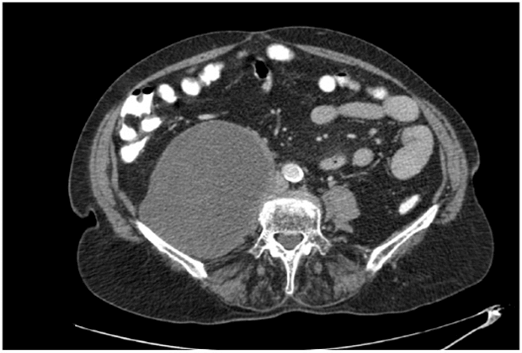
Figure 1: Hematoma of the right psoas.
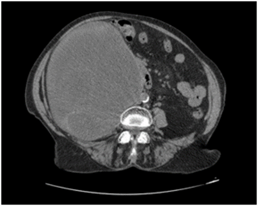
Figure 2: Right retroperitoneal cystic lesion.
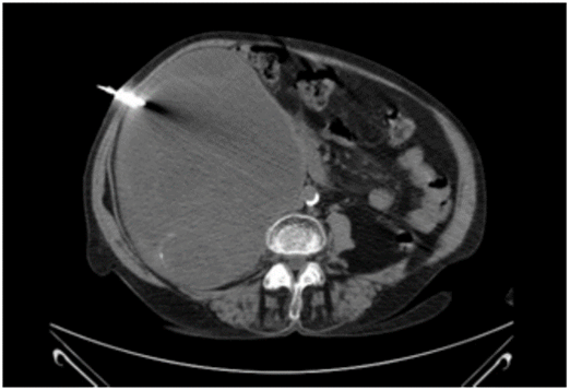
Figure 3: CT-guided biopsy of the lesion.
Article Info
Article Type
Case ReportPublication history
Received: Tue 27, Apr 2021Accepted: Sat 08, May 2021
Published: Tue 25, May 2021
Copyright
© 2023 Lorena Cambeiro Cabré. This is an open-access article distributed under the terms of the Creative Commons Attribution License, which permits unrestricted use, distribution, and reproduction in any medium, provided the original author and source are credited. Hosting by Science Repository.DOI: 10.31487/j.SCR.2021.05.06
Figures & Tables
Table 1: Reported cases of chronic expansive retroperitoneal hematoma.
|
Author |
Patient age (years) |
Patient sex |
Site of the hematoma |
Hematoma size (cm) |
|
Kaneko et al. [1] |
34 |
F |
Above the right kidney |
12 |
|
Hamada et al. [4] |
65 |
M |
At the right iliac fossa |
8 |
|
Irisawa et al.
[5] |
70 |
M |
Below the right kidney |
18 |
|
Yamasaki et al.
[6] |
53 |
M |
Above the left kidney |
12 |
|
Yamada et al. [7] |
59 |
M |
Above the left kidney |
12 |
|
Ried et al. [2] |
79 |
M |
At the right iliac fossa |
NA |
|
Ried et al. [2] |
NA |
NA |
NA |
8 |
|
Syuto et al. [8] |
69 |
M |
Below the left kidney |
20 |
F: female, M: male, NA: data
not available.
References
1. Kaneko G, Shirakawa
H, Kozakai N, Hara S, Nishiyama T et al. (2009) [A case of chronic expanding
hematoma in retroperitoneal space]. Hinyokika Kiyo 55: 603-606. [Crossref]
2. Reid JD, Kommareddi
S, Lankerani M, Park MC (1980) Chronic expanding hematomas. A clinicopathologic
entity. JAMA 244: 2441-2442. [Crossref]
3. Aoki T, Nakata H,
Watanabe H, Maeda H, Toyonaga T et al. (1999) The radiological findings in
chronic expanding hematoma. Skeletal Radiol 28: 396-401. [Crossref]
4. Hamada K, Myoui A, Ueda
T, Higuchi I, Inoue A et al. (2005) FDG-PET imaging for chronic expanding hematoma
in pelvis with massive bone destruction. Skeletal Radiol 34: 807-811. [Crossref]
5. Irisawa M, Tsukuda
S, Amanuma M, Heshiki A, Kuroda I et al. (2005) Chronic expanding hematoma in
the retroperitoneal space: a case report. Radiat Med 23: 116-120. [Crossref]
6. Yamasaki T,
Shirahase T, Hashimura T (2005) Chronic expanding hematoma in the psoas muscle.
Int J Urol 12: 1063-1065. [Crossref]
7. Yamada T, Ishibashi T,
Saito H, Sato A, Matsuhashi T et al. (2003) Case report: chronic expanding hematoma
in the adrenal gland with pathologic correlations. J Comput Assist Tomogr
27: 354-356. [Crossref]
8.
Syuto T, Hatori M, Masashi N, Sekine Y, Suzuki K
(2013) Chronic expanding hematoma in the retroperitoneal space: a case report. BMC
Urol 13: 60. [Crossref]