Atypical presentation of a de Garengeot Hernia: A Case Report
A B S T R A C T
Introduction: An inflamed appendix found within a femoral hernia, a de Garengeot hernia, is a rare occurrence which can present with a variety of different symptoms and be a challenge to surgical management.
Case Presentation: We present the case of a 59-year-old female who presented with migratory right lower quadrant pain and was found to have a de Garengeot hernia diagnosed pre-operatively on CT imaging. She was taken to the OR for an urgent laparoscopic appendectomy with closure of the femoral hernia sack. Given this is not a true repair, the patient was scheduled for an elective femoral hernia repair subsequently.
Discussion: This is the first report of a de Garengeot hernia from the Toronto area. Our case is the first of its kind to present with typical signs of appendicitis without any obvious bulge in the groin.
Conclusion: This case report describes two very common acute surgical presentations, appendicitis and incarcerated hernia, seen simultaneously with only one rare pathology.
Introduction
While abdominal wall hernias are common, affecting up to 5% of the population, femoral hernias are rare, accounting for less than 3% of all groin hernias and are classically seen in women [1]. Migration of the appendix into an incarcerated femoral hernia, termed a de Garengeot hernia, is seen in only 0.9% of femoral hernia cases and is usually discovered incidentally [2]. Subsequent appendicitis within the incarcerated femoral hernia is even more rare with an incidence of 0.08%-0.13% of de Garengeot hernias. Though first described in 1731 by Persian surgeon Rene Jacques Croissant de Garengeot, an ideal guideline for surgical management remains to be elucidated.We describe one of the few reported Canadian cases of this phenomenon.
Presentation of Case
KG, an otherwise healthy 59-year-old female, presented to the emergency department with a one-day history of diffuse abdominal pain which migrated to the right lower quadrant. Pain was worse with right leg flexion and adduction, consistent with a positive obturator sign. Physical examination revealed tenderness over McBurney's point and a positive obturator sign with tenderness over the right groin. A small right groin mass was palpated and there was no overlying erythema. Blood tests showed a normal white blood cell count. CT of the abdomen showed a right femoral hernia containing the distal half of the appendix (Figure 1). The herniated appendix was enlarged to 0.9cm (normal ?0.6cm), with a hyperenhancing wall and surrounding fluid. She was diagnosed with a de Garengeot hernia. Urgent surgery to remove the appendix was advised.
At the time of the laparoscopic operation, the appendix was visualized herniating into the femoral canal medial to the femoral vessels (Figure 2). The appendix was reduced from the femoral canal. Appendectomy was performed. The femoral opening was approximated to trigger an inflammatory response and prevent immediate recurrent herniation while awaiting future hernia repair.
The post-operative period was unremarkable, and the patient was discharged home on postoperative day one. Histology of the appendix showed appendiceal congestion and hemorrhage with reactive mucosal changes consistent with acute appendicitis.
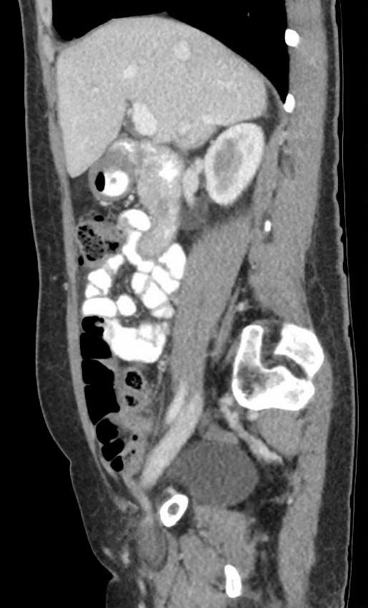
Figure 1: Preoperative CT Scan with arrow pointing toward inflamed appendix within the femoral hernia.

Figure 2: Findings on laparoscopy: Appendix incarcerated in a femoral hernia. Normal appearing appendix proximal to the hernia sac.
Discussion
To our knowledge, we are among the first to report of a de Garengeot hernia in Canada. Our case highlights two common presentations to the emergency room, appendicitis and incarcerated hernia, but within the same patient.
It is currently debated whether, in patients with de Garengeot hernia, the appendicitis occurs before migration into the femoral canal or after the appendix incarcerates into the hernia sac [3]. Our case would support the latter as the portion of appendix outside of the hernia was normal (Figure 2). The same findings were also described on the pre-operative CT imaging. Abnormal attachments of the cecum may increase the risk of migration into a pelvic hernia [3]. Following migration into a femoral hernia, the narrow neck associated with femoral hernia would likely result in extra-luminal compression on the appendix triggering congestion and inflammation.
An established guideline for surgical management of de Garengeot hernias does not exist. The various open approaches to a femoral hernia have been used in the past as they allow adequate visualization of the femoral canal and exposure to the bowel should a resection need to be performed [4]. Given most de Garengeot hernias are diagnosed intraoperatively during an emergency open operation for a strangulated femoral hernia, laparoscopic reduction and repair is unusual [5].
Additionally, most de Garengeot hernias are diagnosed intraoperatively during an emergency operation for a strangulated femoral hernia [6]. Overall pre-operative diagnosis has been shown to be around 14%. Our patients de Garengeot hernia was diagnosed on CT imaging, the gold standard for diagnosing and visualizing the inflamed appendix within the femoral hernia sac [7]. Hence, our case may highlight the importance of CT imaging in the workup of patients with atypical or confusing presentations.
In discussing the surgical approach, a trend to minimally invasive surgery has been seen for acute surgical problems. Laparoscopic technique has been described by Beysens et al. who argue that the appendectomy and hernia repair should be repaired simultaneously with a laparoscopic appendectomy extraperitoneal mesh placement to repair the hernia [9]. Though good results using this approach have been reported, there remains the risk of mesh contamination [10]. In our case, we opted close the peritoneum over the open femoral canal to minimize immediate recurrence and plan for interval hernia repair at a later stage when the patient was symptom free. This approach allowed quick recovery, no surgical site infections and early discharge of the patient.
Conclusion
This case report describes patient with two common acute surgical problems, appendicitis and incarcerated hernia, caused by one pathology. While there are no treatment guidelines, our use of CT for diagnosis as well as minimally invasive surgical approach led to a quick recovery and discharge from hospital.
Funding
None.
Conflict of interest
None.
Article Info
Article Type
Case ReportPublication history
Received: Sat 22, Sep 2018Accepted: Sat 13, Oct 2018
Published: Mon 29, Oct 2018
Copyright
© 2023 Megan Melland-Smith. This is an open-access article distributed under the terms of the Creative Commons Attribution License, which permits unrestricted use, distribution, and reproduction in any medium, provided the original author and source are credited. Hosting by Science Repository.DOI: 10.31487/j.SCR.2018.03.110
Figures & Tables
References
1. Koch A, Edwards A, Haapaniemi S, Nordin P, Kald A (2005) Prospective evaluation of 6895 groin hernia repairs in women. Br J Surg 92: 1553-1558. [Crossref]
2. Theodoros Piperos, Vasileios Kalles, Yousef Al Ahwal, Evangelos Konstantinou, George Skarpas, et al. (2012) Clinical significance of de Garengeot’s hernia: a case of acute appendicitis and review of the literature. Ing J Surg Case Rep 3: 116-117. [Crossref]
3. Khalid Akbari, Claire Wood, Ahmed Hammad, Simon Middleton (2014) De Garengeot’s hernia: our experience of three cases and literature review. BMJ Case Rep. [Crossref]
4. Sorelli PG, El-Marsy NS, Garrett WV (2009) Open femoral hernia repair: one skin incision for all. World J Emerg Surg 4: 44. [Crossref]
5. Jason Ramsingh, Ahmad Ali, Caroline Cameron, Ahmed Al-Ani, Robert Hodnett, et al. (2014) De Garengeot’s hernia: diagnosis and surgical management of a rare type of femoral hernia. J Surg Case Rep. [Crossref]
6. Erdas E, Sias L, Licheri S, Secci L, Aresu S, et al. (2013) De Garengeot hernia with acute appendicitis. G Chir 34: 86-89. [Crossref]
7. Pan CW, Tsao MJ, Su MS (2015) A case of De Garengeot hernia requiring early surgery. BMJ Case Rep. [Crossref]
8. Nguyen ET, Komenaka IK (2004) Strangulated femoral hernia containing a perforated appendix. Can J Surg 47: 68-69. [Crossref]
9. Beysens M, Haeck L, Vindevoghel K (2013) Laparoscopic appendectomy combined with TEP for de garengeot hernia. Case report. Acta chir belg 113: 468-470. [Crossref]
10. Akopian G, Alexander M (2005) De Gerengeot hernia: appendicitis within a femoral hernia. Am surg 71: 526-527. [Crossref]