Journals
Apophysomyces Necrotizing Infection in an Immunocompetent Child
A B S T R A C T
Background: Mucormycoses are uncommon fungal infections with high morbidity and mortality rates. Apophysomyces spp are a known but rare pathogen. Successful treatment necessitates early diagnosis, surgical debridement, and systemic antifungal treatment.
Case Presentation: A 7-year-old boy sustained a minor trauma to his leg and developed necrotizing acute soft tissue infection secondary to Apophysomyces varibilis. He required multiple surgical debridements and systemic antifungal therapy. He recovered well and had excellent results following skin grafting.
Conclusion: Apophysomyces varibilis is an emerging pathogen in immunocompetent patients, most commonly acquired by traumatic innoculation.
Keywords
Apophysomyces, Mucormycosis, necrotizing soft tissue infection.
Abbreviations
ED - emergency department, HD - hospital day, IV - intravenous, WBC - white blood cell, MRI - magnetic resonance imaging , PO - per oral, RLE - right lower extremity.
Background
Mucormycoses are rare infections associated with high rates of morbidity and mortality more commonly seen in immunosuppressed patients but can occur in the immunocompetent [1]. Most cases of mucormycoses are caused by the genus Rhizopus, followed by Mucor and Lichtheimia; together accounting for 70-80% of all cases. Apophysomyces spp, a less common etiology, has been reported in 3% of cases globally [2]. Apophysomyces elegans was first isolated from a soil sample from northern India in 1979; while the first case of mucormycosis secondary to A. elegans in the United States (US) was reported in 1985 [3, 4]. In 2010, three new species of Apophysomyces were identified, including A. varibilis [1]. Successful management of mucormycosis requires early diagnosis, surgical debridement, and systemic antifungal therapy [2]. We herein report a case of an immunocompetent child who developed a necrotizing soft tissue infection from Apophysomyces varibilis following a minor trauma.
Case presentation
A 7-year-old Ethiopian male, with a past medical history significant for typhoid fever and malaria during his first year of life, presented to the emergency department (ED) with a 2-day history of right lower extremity (RLE) pain and swelling. Of note, his parents denied history of foreign travel for the patient in the past 5 years. He had sustained a small laceration to the anterolateral aspect of his right leg secondary to a fall against a concrete step at the beach 5 days prior. Despite local wound care with bacitracin ointment the RLE became progressively edematous. He received one dose of trimethoprim-sulfamethoxazole as an outpatient before becoming febrile to 103°F. At this time, he was given a dose of ceftriaxone and referred to the ED for further care.
On initial exam his temperature was 103F, WBC 9,800, and his leg appeared edematous and indurated without evidence of fluctuance (Fig 1). The presumed cellulitis was initially managed with clindamycin and vancomycin. MRI on hospital day (HD) 1 revealed cellulitis and fasciitis, without necrosis, myositis, or osteomyelitis. On HD 2, as there was some clinical improvement, non-operative management with antibiotics was continued. Cefepime was added for pseudomonal coverage. On HD 6, the development of a fluctuant collection with an associated fever to 104F with a WBC of 16,500 prompted incision and drainage (Figure 2). This revealed infected hematoma which was drained via counter incisions with placement of a Penrose drain. Preliminary results of intra-operative wound cultures were significant for enterococcus, prompting a change in antibiotic regimen to mono-therapy with piperacillin-tazobactam. By HD 8, the patient had continued fevers, for which vancomycin therapy was re-initiated, and noticeably boggy tissue on palpation with a thick, pasty, cheese-like substance expressed on exam. He was subsequently taken for excisional debridement. In the operating room the skin and subcutaneous fat were found to be necrotic, with no necrosis of the underlying fascia and muscle. The following day, preliminary tissue cultures revealed mold, and the patient was started on liposomal amphotericin. Final culture grew Apophysomyces varibilis.
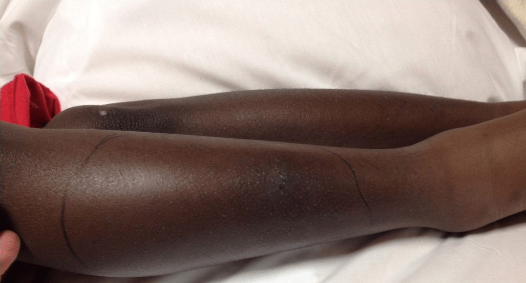
Figure 1: Initial presentation
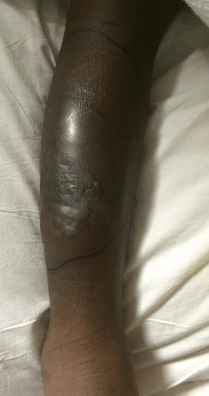
Figure 2: Hospital day 6, prior to first debridement
He was taken for subsequent debridement of devitalized tissue on HD 10. After complete excision and repeat tissue cultures to ensure no residual fungal growth, the wound bed was treated with negative pressure wound therapy. He remained on piperacillin-tazobactam until discharge (HD 17) and was sent home on IV liposomal amphotericin B (Figure 3). He completed 4 weeks of IV liposomal amphotericin B followed by 4 weeks of PO itraconazole. Twenty-one days after initial presentation to the ED he underwent split thickness skin graft with 100% take (Figure 4) He healed well and had no recurrence of infection.

Figure 3: Wound appearance at time of discharge.

Figure 4: Healed skin graft.
Discussion
Mucormycoses are rare infections that more commonly afflict immunocompromised patients; however, recent reports demonstrate that Apophysomyces spp are an emerging pathogen in the immunocompetent host [5]. As seen in our case report, Apophysomyces varibilis has specifically been noted to mainly affect immunocompetent patients [1]. Traumatic inoculation of the organism is the most common mode of Apophysomyces infections, with almost half of all documented cases secondary to trauma. The most common sites are cutaneous/subcutaneous (53%), with local invasion and necrotizing soft tissue infections a common feature (83%), and the majority (79%) progressing to necrotizing fasciitis or gangrene. Similar to our patient, cutaneous infections can manifest with pain, swelling, abscesses, ulceration and necrosis, with the most common locations being the extremities, abdominal wall and perineum [2].
Management of these patients require both medical and surgical treatment. The antifungals that are standard of care for the management of Apophysomyces mucormycosis are amphotericin B and itraconazole, with recent studies showing promising results for other therapeutic agents [6]. Surgical debridement is associated with a decrease in fungal load, as well as improved tissue penetration of antifungal agents post resection. A prospective study demonstrated that the survival rate of amphotericin B plus surgery was 61.5% compared to 10.3% for amphotericin B alone (OR = 0.2, p < 0.04) [7]. In our case report, aggressive surgical debridement was initially delayed due to MRI findings not consistent with necrotizing infection and initial cultures demonstrating a bacterial source. Due to progression of the infection with initial treatment and cultures demonstrating mold, he underwent aggressive surgical debridement and IV antifungal therapy.
Conclusion
In conclusion, patients presenting with a necrotizing acute soft tissue infection, with or without a history of trauma, mucormycosis must be considered in the differential, even in the immunocompetent. In those suspected to have mucromycosis, early diagnosis and treatment with aggressive surgical debridement and systemic antifungal therapy is required for patient survival.
Ethics approval and consent to participate
No IRB approval required for this case report.
Consent for publication
Written consent for publication obtained from the patient’s guardians.
Availability of data and materials
No data set used for this article
Conflict of interest
The authors have no conflicts of interest.
Funding
No sources of funding.
Authors Contributions
Each author has made substantial contributions to the information or material submitted for publication.
Article Info
Article Type
Case ReportPublication history
Received: Tue 15, Jan 2019Accepted: Mon 11, Feb 2019
Published: Thu 07, Mar 2019
Copyright
© 2023 Shannon Longshore. This is an open-access article distributed under the terms of the Creative Commons Attribution License, which permits unrestricted use, distribution, and reproduction in any medium, provided the original author and source are credited. Hosting by Science Repository.DOI: 10.31487/j.SCR.2019.01.002
Author Info
Corresponding Author
Shannon LongshoreDivision of Pediatric Surgery, Department of Surgery, Brody School of Medicine at East Carolina University, 600 Moye Blvd MA 207, Greenville, NC, 27834 USA
Figures & Tables
References
- Rodriguez JY, Morales-Lopez SE, Rodriguez GJ, Álvarez-Moreno CA, Ocampo W, et al. (2017) Necrotizing fasciitis caused by Apophysomyces variabilis in an immunocompetent patient. Med Mycol Case Rep 20: 4-6. [Crossref]
- Gomes MZ, Lewis RE, Kontoyiannis DP (2011) Mucormycosis caused by unusual mucormycetes, non-Rhizopus, -Mucor, and -Lichtheimia species. Clin Microbiol Rev 24: 411-445.
- Misra PCS, K J, Lata K (1979) Apophysomyces, a new genus of the Mucorales. Mycotaxon.
- Wieden MA, Steinbronn KK, Padhye AA, Ajello L, Chandler FW (1985) Zygomycosis caused by Apophysomyces elegans. J Clin Microbiol 22: 522-526. [Crossref]
- Meis JF, Chakrabarti A (2009) Changing epidemiology of an emerging infection: zygomycosis. Clin Microbiol Infect 5: 10-14. [Crossref]
- Prakash H, Ghosh AK, Rudramurthy SM, Paul RA, Gupta S, et al. The environmental source of emerging Apophysomyces variabilis infection in India. Med Mycol 54: 567-575. [Crossref]
- Bala K, Chander J, Handa U, Punia RS, Attri AK (2015) A prospective study of mucormycosis in north India: experience from a tertiary care hospital. Med Mycol 53: 248-257. [Crossref]