
Journals
Anterior Hollow Screw Fixation Combined with Posterior Lateral Mass Screws Fixation to Treat A Huge Tear Drop Fracture of The Axis
A B S T R A C T
Teardrop fracture of axis is rarely seen, especially the huge type. The surgical approach is controversial and individualized in the literature. We reported a special case to share our experience of anterior hollow screw fixation combined with posterior unilateral lateral mass screws fixation to treat a huge teardrop fracture of the axis (HTDFA). A 42-year-old male was referred to our department; he presented neck pain after a vehicle accident. Neck movement was limited with no neurological compromise. Cervical x-rays showed a huge tear drop fracture of the anterior–inferior corner of the axis,the lower cervical spine degeneration and poor cervical curvature. Cervical CT confirmed the avulsed ratio of inferior endplate of axis up to 62%. Cervical MRI showed the anterior longitudinal ligament disruption associated with disk injury and instability of C2-3. In order to save the motion segment of C2-3 and decrease the possibility of lower cervical degeneration if C2-3 fusion occurs, a surgical plan involving anterior reduction, hollow screw fixation combined with posterior unilateral mass screws fixation was performed. The 3 months postoperative x-ray and CT scan showed the tear drop fragment had completely healed. The internal fixations were removed timely for 6 months after surgery. The patient had an excellent function of cervical spine without any clinical symptoms. The 12 months postoperative X-ray showed radiographic union of the bony fragment and a good motion range without any instability. We conclude that anterior hollow screw fixation combined with posterior unilateral lateral mass screws fixation can be the treatment of choice for HTDFA.
Keywords
Teardrop fracture, axis, anterior surgical procedures, posterior surgical procedures
Introduction
Teardrop fracture of the axis which originally described by Schneider and Kahn is extremely rare spinal fracture [1]. It is defined as a displaced triangular fragment at the anterior-inferior corner of the axis caused by various types of trauma. Due to questioning the stability of this injury by certain authors, the treatment strategy of teardrop fracture of the axis remains controversial and individualized [2]. However, for a huge and significant displaced fragment, especially combined with intervertebral disc injury and/or instability of C2-3, almost all authors chose C2/3 fusion surgery regardless of anterior or posterior approach in previous studies [3-9]. Almost no literature has been reported about no-fusion surgery method to treat huge teardrop fracture of the axis [HTDFA] with intervertebral disc injury and/or instability of C2-3. In this study, we described the successful treatment of a patient of HTDFA with intervertebral disc injury and instability of C2-3 by non-fusion method via anterior-posterior approach.
Case Report
A 42-year-old gentleman presented to author’s department with neck pain and mild dysphagia 2 days after a vehicle accident. He crashed with a car while riding a motorcycle. He struck his head and face on the ground when fell. He had not received any special treatment except for the protection of soft collar. Physical examination showed cervical muscle spasm and limitation of neck motion. His neurologic examination revealed no abnormality. Cervical x-rays revealed a huge avulsed and rotated fragment from the anterior-inferior angle of the axis on the lateral radiograph. There was a slight retrolisthesis of the body of the axis. And the lower cervical spine showed degeneration and poor curvature (Figure 1).
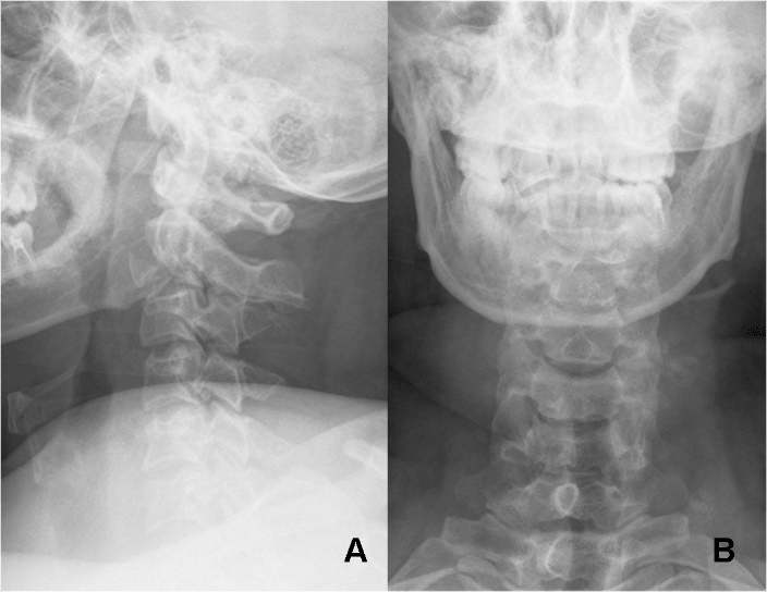
Figure 1: Preoperative anteroposterior and lateral x-rays revealed huge tear drop fracture of anterior–inferior corner of the axis.
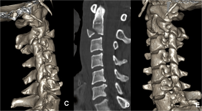
Figure 2: Preoperative cervical CT confirmed huge tear drop fracture of anterior-inferior corner of the axis, and the discontinuity of the cortex.
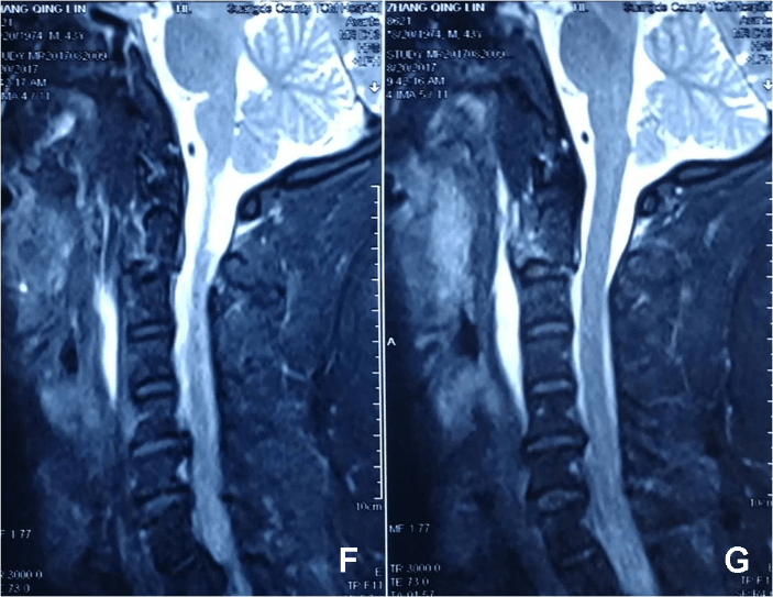
Figure 3: Preoperative cervical MRI showed hyperintensity in C2/3 intervertebral disc and pre-cervical soft tissue.
Cervical CT scan confirmed the fracture line of fragment traversed the lower endplate of the body of axis completely with the avulsed ratio of inferior endplate of axis up to 62% by using the Tao 's method [8]. And the huge tear drop fragment had a significant displacement (Figure 2). Cervical MRI showed the tear drop fracture and disruption of the anterior longitudinal ligament associated with C2-3 disk injury and instability of C2-3. Prevertebral soft-tissue (PVST) swelling for this patient was diffuse (Figure 3). After reviewing the literature, we found that only a dozen cases of HTDFA were reported in previous studies and the methods of operation were almost adopted C2-3 fusion surgery [3-9]. Considering the patient’s age (42y) and his significant degeneration of C5/6, C6/7 and poor cervical curvature, C2-3 fusion surgery might accelerate the lower cervical degeneration. Hence, a surgical plan involving anterior reduction, hollow screw fixation combined with posterior unilateral mass screws fixation via anterior-posterior approach was planned for saving the motion segment of C2-3.
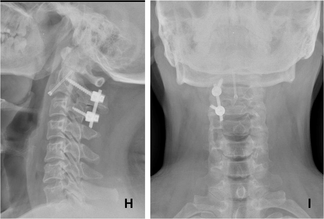
Figure 4: Three months postoperative X-ray showed the tear drop fragment healing with a slight local angle deformity and rotated deformity.
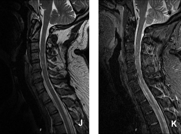
Figure 5: The postoperative T2-MRI after the internal fixations being removed showed the intervertebral disk of C2/3 had completely repair with normal signal.
The surgery was performed using a classical posterior approach firstly, and then anterior approach by an experienced surgeon after transnasal induction of general anesthesia on 31 August 2017. One day postoperative x-ray showed a good reduction of the huge tear drop fracture fragment with a slight rotated deformity. His dysphagia disappeared 1 week after operation. The patient was instructed to wear a cervical collar until their return to the department for a follow-up examination 1 months after their surgery. The 3 months postoperative X-ray showed the tear drop fragment had completely healed with a slight local angle deformity and rotated deformity (Figure 4). The internal fixations were removed timely for 6 months after surgery for preservation of the C2/3 intervertebral disc function. The postoperative T2-MRI after the internal fixations being removed showed the intervertebral disk of C2/3 had completely repair with normal signal (Figure 5). At the final follow-up of 12 month, the patient had an excellent function of cervical spine without any clinical symptoms (no neurological deficits, no stiffness or tenderness over the spinal process of the axis) .And the X-ray showed radiographic union of the bony fragment with good curvature (Figure 6).
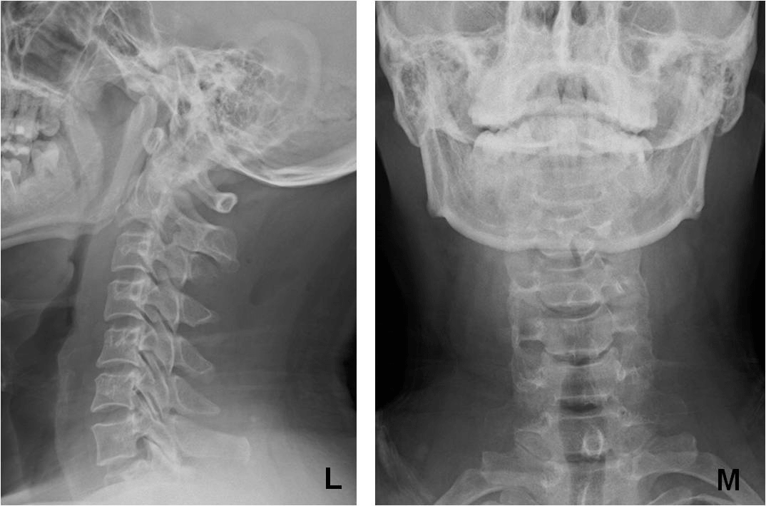
Figure 6: The X-ray one year later showed radiographic union of the bony fragment with good curvature.
Discussion
Generally, it is often regarded as a huge tear drop fracture if the avulsed ratio of inferior endplate of axis is above 50% [3, 7]. In this patient, the fracture line lies up to 58% of lower endplate of the axis in the sagittal plane. The injury mechanism of tear drop fracture of the axis is generally considered hyperextension. The forehead wound in this patient suggested a direct blow leading to an extension moment of the cervical spine. When the hyperextension of the cervical spine occurs, the odontoid process of the axis acts as a moment arm of leverage to transfer the backward stress, the posterior elements of axis acts as a fulcrum. At this moment, extension of the anterior longitudinal ligament could cause the avulsion fracture at anterior inferior portion of the axis body and various degrees of injury of the anterior longitudinal ligament [9-10]. The hyperextension has also leaded to intervertebral disc injury and C2/3 instability in this patient.
The huge tear avulsed fragment usually had a significant displacement and rotation in most patients. There is lots of controversy over the conservative and surgical methods because both seems to be effective in the treatment of HTDFA. Due to the lack of reducement completely with skull traction or cervical collar, the fracture bone fragment usually had healed with significant malunion when choosing conservative management [10]. Furthermore, the HTDFA could cause the compression of esophagus and induce the dysphagia during conservative treatment in some patients [11]. Hu et al. retrospectively reviewed 16 patients with tear drop fracture of the axis and considered that large fragment size, displacement or angulation, intervertebral disc injury, neurologic deficit, or signs of instability were reasonable indications for surgical treatment [5]. Regardless of anterior or posterior approach, the surgical strategy should be aimed at restoring stability and cervical alignment by adopting C2/3 fusion in previous papers. However, the reason why we applied C2/3 non-fusion method was as follows: The lower cervical spine has been obviously degenerated with poor alignment although the patient was only in his forties. If C2/3 fusion, it may accelerate the degeneration of lower cervical spine. It was reported that the damage to the anterior longitudinal ligament and intervertebral disc were significantly associated with initial cervical segmental instability [12]. We think the main factors of C2/3 instability are the huge avulsed fracture, the rupture anterior longitudinal ligament and the rupture of the intervertebral disc. The anterior hollow screw fixation can reconstruct the integrality of the bone and ligament. If the ruptured intervertebral disc heals well, the physiological function and stability of C2/3 can be retained. Compared with external fixation, such as a halo vest or skull traction or cervical collar, short-term internal fixation can provide greater possibility for intervertebral disc healing by better restrictions the intervertebral movement of C2/3.
The high anterior cervical retropharyngeal approach is the most commonly used to perform the surgery in the previous studies [9]. However, the high anterior cervical spine involves a lot of complex anatomic structures, such as the Carotid sheath, hypoglossal nerve, superior laryngeal nerve. The occurrence of hypoglossal nerve and superior laryngeal nerve injury during operation can lead to a temporary or permanent dysphagia [13-14]. Park et al. reported that among fifteen patients whose upper cervical surgery were performed via the perivascular extraoral retropharyngeal approach, 1 patient developed permanent dysphagia related to the hypoglossal nerve and 2 had transient dysphagia [15]. We used the routinely approach of the axis odontoid screw fixation. The location of the incision is at the level of C4/5. The incision can quickly finish the avulsed fragment reduction and fixation while avoiding these important nerve structure.
In addition to restricting the intervertebral movement of C2/3, the other aim of the lateral mass screw fixation is to assist the anterior hollow screw fixation. As we know, the cervical position of the anterior approach needs to bend back for exposing the nail point of the fracture fragment well. However, the excessive bending of the neck will result in the greater separation of bone mass and more difficulty of the fracture fragment reduction. We first completed the lateral mass screw fixation and then turned over for anterior operation. The immovable intervertebral space limited by the fixed lateral mass screw would not leading to the increasing displacement of bone mass and be available for fracture fragment reduction and fixation during anterior surgery. It is considered that prolonged fixation of C2/3 might lead to degeneration of intervertebral disc tissue. We took out the internal fixator without any delay six month after operation. The MRI of 6 months post-operation confirmed that the C2/3 disc had healed completely with normal signal. This indicated that the intervertebral disk had not degenerated in condition of posterior fixation of C2–3. Moreover, the patient had an excellent function of cervical spine without any clinical symptoms at the final follow-up of 1year. Therefore, we think that anterior hollow screw fixation combined with posterior unilateral lateral mass screws can be a good choice for HTDFA
Acknowledgments and funding
This work was supported by 1112 talent project of Huzhou and New medical youngster project of Zhejiang and Huzhou Science and Technology Project (2017GY38) and Medical Key Points of Nanjing Military Region (15ZD003).
Declaration of interest
The manuscript submitted does not contain information about medical device(s)/drug(s). The authors declare that they have no conflicts of interest concerning this article.
Article Info
Article Type
Case ReportPublication history
Received: Sat 08, Jun 2019Accepted: Fri 21, Jun 2019
Published: Sat 20, Jul 2019
Copyright
© 2023 Huang Fei. This is an open-access article distributed under the terms of the Creative Commons Attribution License, which permits unrestricted use, distribution, and reproduction in any medium, provided the original author and source are credited. Hosting by Science Repository.DOI: 10.31487/j.SCR.2019.03.13
Author Info
Corresponding Author
Huang FeiDepartment of Orthopaedic Surgery, No. 98 Hospital of PLA, Huzhou, China
Figures & Tables
References
- Schneider RC, Kahn EA (1956) Chronic neurological sequelae of acute trauma to the spine and spinal cord. I. The significance of the acute-flexion or tear-drop fracture-dislocation of the cervical spine. J Bone Joint Surg Am 38-A: 985-997. [Crossref]
- Bauer DR, Errico TJ (1991) Cervical Spine Injuries. Spinal Trauma. Philadelphia: J. B. Lippincott 71-121.
- Yang X, Zheng B, Hao D, Liu B, Yan L et al. (2015) Treating huge Tear-Drop Fracture of Axis With Trapezoidal Bone: A Case Report and Literature Review. Spine 40: E1187-E1190. [Crossref]
- Vialle R, Schmider L, Levassor N, Rillardon L, Drain O et al. (2004) Extension tear-drop fracture of the axis: a surgically treated case. Rev Chir Orthop Reparatrice Appar Mot 90: 152-155. [Crossref]
- Hu Y, Kepler CK, Albert TJ, Hann S, Ma WH et al. (2016) Conservative and Operative Treatment in Extension Teardrop Fractures of the Axis. Cin Spine Surg 29: E49-E54. [Crossref]
- Xu G, Li W, Bao G, Sun Y, Wang L et al. (2014) Tear-drop fracture of the axis in a child with an 8 year follow-up: a case report. J Pediatr Orthop B 23: 299-305. [Crossref]
- (2014) with an 8-year follow-up: a case report. J Pediatr Orthop B 23:299-305.
- Jiang T, Yin H, Ren, XJ, Chu TW, Wang WD et al. (2017) Anterior reduction and fusion for treatment of massive tear drop fracture of axis combining with inferior endplate serious traversed lesion: A retrospective study. J Orthop Sci 22: 816-821. [Crossref]
- Deniz FE, Cagli S, Zileli M (2007) Compressive hyperextension injury of C2-C3 managed with anterior plate fixation: case report. Turk Neurosurg 17: 125-128. [Crossref]
- Ma L, Yang Y, Gong Q, Ding C, Liu H et al. (2016) Anterior Reduction, Discectomy, and Three Cortical Iliac Bone Grafting With Instrumentation to Treat A Huge TearDrop Fracture of the Axis: A Case Report and Literature Review. Medicine 95: e3376. [Crossref]
- Korres DS, Zoubos AB, Kavadias K, Babis GS, Balalis K et al. (1994) The ‘‘tear drop’’ (or avulsed) fracture of the anterior inferior angle of the axis. Eur Spine J 3: 151-154. [Crossref]
- Watanabe M, Sakai D, Yamamoto Y, Sato M, Mochida J et al. (2011) Clinical features of the extension teardrop fracture of the axis: review of 13 cases. J Neurosurg Spine 14: 710-714. [Crossref]
- Maeda T, Ueta T, Mori E, Yugue I, Kawano O et al. (2012) Soft-tissue damage and segmental instability in adult patients with cervical spinal cord injury without major bone injury. Spine 37: E1560-E1566. [Crossref]
- Yu Fengbin, Wang Xinwei , Yang Haisong, Chen Yu, Lia Xiaowei et al. (2013) Dysphagia after anterior cervical discectomy and fusion: a prospective study comparing two anterior surgical approaches. Eur Spine J 22: 1147-1151. [Crossref]
- Hur H, Lee JK, Jang JW, Kim TS, Kim SH (2014) Is it feasible to treat unstable hangman's fracture via the primary standard anterior retropharyngeal approach? Eur Spine J 23: 1641-1647. [Crossref]
- Park SH, Sung JK, Lee SH, Park J, Hwang JW et al. (2007) High anterior cervical approach to the upper cervical spine. Surg Neurol 68: 519-524. [Crossref]