Journals
Amyand's hernia: a mini review and two case reports
A B S T R A C T
Claudius Amyand was the first to perform a successful appendicectomy in 1735 as well as describe an ‘Amyand’s Hernia’. It is an uncommon condition, in which an inguinal hernia, a protrusion of abdominal contents through the inguinal canal, contains an incarcerated vermiform appendix, irrespective of whether it is inflamed or not. The reported incidence is less than 1.7% with histologically normal appendix and less than 0.1% having an inflamed or perforated appendix. We report a case of acute appendicitis after its incarceration in the inguinal hernia and another case of Amyand’s hernia containing a healthy, non-inflamed appendix. Comorbidities, clinical presentation, investigations as well as surgical management are presented. One patient has provided us with a written consent for image disclosure. Both patients presented with signs and symptoms of incarcerated irreducible inguinal hernia. Contrast computer tomography (CT) was the only modality to diagnose the hernia sac contents preoperatively. A laparoscopic repair was performed on the first case (inflamed appendix) and an open repair was chosen for the second case (non-inflamed). Both patients had an uncomplicated and uneventful postoperative recovery.
Amyand hernia is a rare entity with variable presentations; from a reducible inguinal hernia containing a normal appendix, to acute abdomen due to perforation of acute appendicitis secondary to incarceration. Only imaging can verify the contents of an incarcerated inguinal hernia and the approach varies upon those findings. It is generally accepted that surgical treatment involves hernia repair with or without concomitant appendicectomy
Keywords
Amyand hernia, vermiform appendix, appendicectomy, hernia repair, open vs laparoscop
Introduction
An Amyand’s hernia is a rarely encountered type of hernia, characterised by the appendix becoming incarcerated within an inguinal hernia, irrespective of whether the appendix itself is normal, inflamed or exhibiting any other pathology. This phenomenon was first described by Claudius Amyand (1660–1740), a French surgeon primarily known for performing the first appendicectomy successfully on a 1year-old boy in 1735 [1]. More specifically, he described a perforated appendix in an inguinal hernia sac. This pathology is an even rarer variation of an Amyand’s hernia [2]. Overall, an AH has been described in less than 1.7% of hernias in the literature thus far, with the cases of an inflamed or perforated incarcerated appendix making up only about 0.1% of the cases. In addition, cases have been described in all age groups but have been more commonly encountered in children. This is most probably due to a patent processus vaginalis predisposing to this condition [3]. The importance of recognising this condition lies in the fact that a variable, yet significant, mortality has been associated with it (5.5%–30%), mostly due to peritonitis and sepsis [4]. Complications from an inflamed or perforated appendix contained in an Amyand’s hernia can extend to involve the right testicle and associated structures, form intra-abdominal abscesses or even cause necrotising fasciitis of the inguinal region or anterior abdominal wall [5]. An Amyand’s Hernia poses a diagnostic challenge, as its identification is often incidental, on imaging or intra-operatively [6, 7, 8, 9]. This is because clinically, it can present with variable, non-specific signs and symptoms or be mistaken for a simple incarcerated inguinal hernia. When the pre-op differential diagnosis is that of a strangulated hernia, imaging can often be by-passed, and the diagnosis occurs intra-operatively. However, in cases in which the presentation can be more complex and intra-abdominal pathology is suspected, imaging is used, most commonly a CT scan, and thus a pre-operative diagnosis is feasible [5].
We report a case of acute appendicitis brought on by its incarceration in the inguinal hernia, diagnosed intra-operatively, and another case of Amyand’s hernia containing a healthy, non-inflamed appendix identified pre-operatively. In this paper we support the fact that an Amyand’s hernia can present with a range of signs and symptoms, depending on the appendiceal pathology within. More specifically, an AH can contain a normal, asymptomatic appendix or an abscess formed secondary to perforation of an acutely inflamed appendix. Because of the rarity of this type of hernia, a standard management approach has not been established, however, thus far it has been described that the most common surgical management is that of a hernia repair with or without concomitant appendicectomy [10].
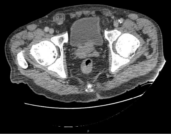

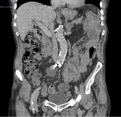
Figure 1, 2, 3: 86year old male with Right Inguinal Hernia (RIH) containing a non-inflamed appendix and ceacum, known as Amyand hernia (Case 2)
Case Report 1
A 58year old female presented to the emergency department with severe abdominal pain and a palpable, tender and non-reducible right inguinal hernia (RIH) with signs of inflammation. Her previous medical history included hypertension and asthma, but no previous abdominal surgeries. Her vital signs on arrival were BP 143/105mmHg, HR 110bpm and temperature 36.20C. Blood results included a White Blood Cell count (WBC) 11.3X10⁹/L and C-Reactive Protein (CRP) 192.2 mg/L, supporting the clinical diagnosis of hernia incarceration and inflammation.
The patient was clinically diagnosed as having an incarcerated femoral hernia and the decision for emergency surgical intervention was made at this stage, without any imaging requirement. She was taken to theatre within two hours of admission and a laparoscopy was performed. The intraoperative findings contradicted the initial diagnosis, revealing an incarcerated RIH containing an inflamed appendix and an incarcerated loop of small bowel (SB) causing obstruction and ischaemia. An intraoperative diagnosis of an Amyand hernia was made. The patient underwent a laparoscopic appendicectomy, small bowel resection and anastomosis as well as transabdominal pre-peritoneal (TAPP) repair of the right inguinal hernia with mesh. Histology report showed necrotic and inflamed small bowel and appendix. Her postoperative course was unremarkable.
Case Report 2
An 86year old male presented to the emergency department and was admitted with severe right inguinal pain and a known long standing RIH. His vital signs were stable and blood results normal. Previous medical history included hypertension, rheumatoid arthritis but no surgical history. A partially-reducible right groin mass with tenderness on palpation was noted on physical examination. The inguinal hernia was reduced partially on admission, but the abdominal x-ray revealed dilated small bowel loops. A CT scan of the abdomen and pelvis with oral enhancing contrast was performed and showed a RIH with an incarcerated appendix and ceacum but the absence of any signs of inflammation (Figures 1, 2, 3). A preoperative diagnosis of Amyand’s hernia was made and the patient underwent an open RIH mesh repair under general anaesthesia with no resection of the bowel or the appendix. His postoperative course was also unremarkable.
Discussion
An Amyand’s Hernia is defined as an inguinal hernia containing an incarcerated appendix [2]. This is a rarely encountered entity since, more commonly, an inguinal hernia will contain small or large bowel, or only omentum. Less common, and often discovered intra-operatively, inguinal hernia contents can be unexpected such as in the case of a Meckel’s diverticulum (Littre’s hernia), a segment of the bowel’s circumference (Richter’s hernia) or even a portion of the bladder (sliding hernia) [5].
The incidence of an Amyand’s hernia is low according to reported cases to this day (0.19-1.7%) but becomes even lower when it contains a pathological appendix i.e. one that is inflamed (0.07-0.13%) or complicated by perforation and abscess formation (0.1%) [4, 5, 10]. In terms of the cases reported in the literature thus far, the age of patients who have had an AH varies greatly, and ranges from 19 days old to 89 years old [5, 10]. This type of hernia is more frequent in men however, inguinal hernias are overall more frequent in males [10]. AHs are more frequently found on the right, possibly due to the anatomical position of the appendix as well as the fact that right inguinal hernias tend to be more common. A few cases of left sided Amyand hernias have been reported, and in those cases, it is important to consider an associated condition which would enable such a variation, including situs inversus, bowel malrotation or increased mobility of the caecum [11]. Only a handful of cases have been reported so far where an AH was complicated by an appendiceal carcinoma [12-14]. Over the course of three years at our institute (November 2015 to November 2018), we had a case of a 58year old female with incarceration, inflammation and perforation of the appendix within the right inguinal hernia sac (Case 1) and an 86year old male with non-inflamed appendix contained within the hernia sac of a non-reducible right inguinal hernia (Case 2).
Both cases presented to the emergency department with ‘acute abdomen’. Case 1 presented with signs of an incarcerated right femoral hernia, hence this is the most frequent diagnosis in females, whereas Case 2 presented with signs of an irreducible right inguinal hernia. Accurate pre-operative diagnosis became possible in the second case. The patient’s advanced age and abdominal x-ray, demonstrating dilated small bowel loops, raised clinical suspicion of a mass as a cause for his symptoms and bowel obstruction. Hence, a CT scan with intravenous contrast was obtained. The younger, female patient (Case 1) had a preoperative differential diagnosis of incarcerated femoral hernia based on laboratory results and clinical examination, suggesting inflammation, strangulation and possible perforation. Therefore, decision was made for emergency surgery without any imaging.
Pre-operative diagnosis of Amyand's hernia can be difficult and uncommon [15]. Up until 1999, only 1 out of 60 cases had been successfully diagnosed as AH prior any intervention [16]. Clinical examination of an AH reveals non-specific signs of hernia pathology, more specifically those elicited in patients with an inguinal or femoral hernia. Laboratory findings, just like in other incarcerated hernias, can range from normal or low-grade inflammation to full systemic sepsis response, but again would not help in identifying the mechanism or type of intra-abdominal processes occuring. The pathophysiology of an inflamed appendix and its sequlae within an inguinal hernia has been a matter of controversy in the literature. Due to the rarity of the condition as well as the limited instances of its diagnosis pre-operatively, it has been challenging to outline clearly the course of events leading up to intra-abdominal infection. The most accepted theory is that the appendix herniates into the sac, and a combination of micro-trauma, fibrosis, muscle contractions and raised intra-abdominal pressures results in its swelling. Subsequently, the appendix becomes incarcerated, impairing its own blood supply and promoting bacterial overgrowth in the area. This will result in inflammation and, if not recognised and treated at that stage, perforation, intra-abdominal infection and/or abscess formation [16, 17].
An Amyand hernia is an inguinal hernia containing the appendix within the hernia sac. The appendix can become incarcerated within the hernia sac and thus becomes vulnerable to trauma and adhesions. It is still unknown whether the appendix position within the hernia sac makes it more prone to inflammation as well as whether it increases the chances of the hernia becoming incarcerated. In other words, whether the anatomical variation which gives rise to an AH predisposes to pathology in the area. Once the appendix is incarcerated, it is restricted from sliding back into the abdominal cavity and is subsequently associated with an increased risk of inflammation. When the appendix that has herniated through an inguinal hernia does not exhibit any inflammatory changes or other pathology, it can be retracted into the peritoneal cavity. Whether it is removed or not is a matter of controversy. On one hand, some surgeons support to avoid an appendicectomy is to avoid any potential contamination of the surgical field and thus minimise the chances of post-operative infection [18, 5]. It has also been argued that an appendicectomy in children might have a negative effect on the immune response, as well as limit any of its potential use in future interventions (such as for urinary diversions or reconstruction procedures) [3, 5].
On the other hand, if an appendicectomy occurs concomitantly by a highly skilled surgeon, the rate of intra-abdominal contamination will be lower. The benefits of removing the appendix arise mainly from prevention of future appendiceal inflammation and associated complications [19]. This is in fact heavily supported by data reporting lower rates in morbidity after appendicectomy of a normal appendix in an AH when compared to the alternative approach i.e. retracting the healthy appendix into the abdominal cavity. As suggested by Ofili, the retraction and manipulation of the appendix from the hernia might be sufficient to elicit an inflammatory process in the future [20]. In fact, 2 cases of acute appendicitis have been reported following inguinal hernia repair without incidental appendicectomy, and 11 cases of herniorrhaphy with incidental appendicectomies without any wound infection or hernia recurrence [1]. The use of mesh to repair the hernia has also been a topic of controversy. Some see it as a contraindication due to the increased chances of having an inflammatory response from the contaminated abdominal wall and the synthetic prosthesis [21, 22]. Sharma et al. argue that it is safe to retain a normal appendix and to use mesh for the hernia repair [8].
When an inflamed appendix is encountered, a laparoscopic approach can be employed to retract the appendix back into the peritoneal cavity and subsequently perform an appendicectomy and an open hernia repair [23]. Over recent years, there has been an increase in laparoscopic approaches for suspected appendicitis and associated complications of an AH [5]. The alternative approach, used when there is suspected intra-abdominal or pelvic infection and abscess formation, is that of a lower midline laparotomy [8, 24]. Solecki et al. recommended lower midline laparotomy, and Shouldice’s herniorrhaphy in cases of gangrenous acute appendicitis in AH [24]. Our surgical approach included hernia repair with mesh in both cases and then ranged from laparoscopic appendicectomy for appendicitis to reducing a healthy appendix intra-abdominally. Both patients had an uneventful recovery without any post-operative complications recorded to date.
Conclusions
An Amyand hernia is a rare entity with varying presentations; from a reducible inguinal hernia containing a normal appendix to acute abdomen due to perforation of acute appendicitis secondary to incarceration. Only imaging can verify the contents of an incarcerated inguinal hernia and the approach varies upon those findings. It is generally accepted that surgical treatment includes a hernia repair with or without concomitant appendicectomy.
Funding
This research did not receive any specific grant from any funding agency in the public, commercial, or not-for-profit sector.
Compliance with ethical standards
Informed consent: Obtained from one patient for disclosing figures
Disclosure
Sylvia Krivan, Andrea Giorga and Omer Al-taan have no conflicts of interest or financial ties to disclose.
Article Info
Article Type
Case ReportPublication history
Received: Thu 07, Mar 2019Accepted: Wed 27, Mar 2019
Published: Wed 03, Apr 2019
Copyright
© 2023 Sylvia Krivan. This is an open-access article distributed under the terms of the Creative Commons Attribution License, which permits unrestricted use, distribution, and reproduction in any medium, provided the original author and source are credited. Hosting by Science Repository.DOI: 10.31487/j.SCR.2019.02.013
Author Info
Corresponding Author
Sylvia KrivanUpper Gastrointestinal and Bariatric unit, Luton and Dunstable University Hospital, United Kingdom
Figures & Tables
References
- Eduardo Smith-Singares, Joseph Adjei Boachie, Izaskun Melania Iglesias (2016) A rare case of appendicitis incarcerated in an inguinal hernia. J Surg Case Rep 6: rjw096. [Crossref]
- Eric Paul Ebaugh, Kara Hessel, Kahdi Udobi (2016) Appendiceal perforation, necrotizing groin infection and spermatic cord necrosis in a case of Amyand’s hernia. Int J Surg Case Rep. 24: 172-174. [Crossref]
- Baldassarre E, Centozea A, Mazzei A, Rubino R (2009) Amyand’s hernia in premature twins. Hernia 13: 229-30. [Crossref]
- D’Alia C, Lo Schiavo MG, Tonante A, Taranto F, Gagliano E et al. (2003) Amyand’s hernia: case report and review of the literature. Hernia 7: 89-91. [Crossref]
- Ivashchuk G, Cesmebasi A, Sorenson EP, Blaak C, Loukas M et al. (2014) Amyand’s hernia: A review. Med Sci Monit 20: 140-146. [Crossref]
- Ali SM, Malik KA, Al-Qadhi H (2012) Amyand’s hernia: study of four cases and literature review. Sultan Qaboos Univ Med J 12: 232-236. [Crossref]
- R Hutchinson (1993) Amyand’s hernia. J R Soc Med 86: 104-105. [Crossref]
- Sharma H, Gupta A, Shekhawat NS, Memon B, Memon MA (2007) Amyand’s hernia: a report of 18 consecutive patients over a 15-year period. Hernia 11: 31-35. [Crossref]
- Lyass S, Kim A, Bauer J (1997) Perforated appendicitis within an inguinal hernia: Case report and review of the literature. Am J Gastroenterol 92: 700-702. [Crossref]
- Kose, Emin, Abdullah Sisik, Mustafa Hasbahceci (2017) Mesh Inguinal Hernia Repair and Appendectomy in the Treatment of Amyand’s Hernia with Non-Inflamed Appedices. Surgery Research and Practice 7696385.
- Al-Mayoof AF, Al-Ani BH (2014) Left-Sided amyand Hernia: Report of two cases with review of literature. European J Pediatr Surg Rep 2: 63-66. [Crossref]
- Ioannis Karanikas, Argyrios Ioannidis, Petros Siaperas, Georgios Efstathiou, Ioannis Drikos et al. (2015) Incarcerated Amyand hernia with simultaneous rupture of an adenocarcinoma is an inguinal hernia sac: a case report. J Med Case Rep 9: 120. [Crossref]
- Wu CL, Yu CC (2010) Amyand's hernia with Adenocarcinoid tumor. Hernia 14: 423-425. [Crossref]
- Gregorios Christodoulidis, Konstantinos Perivoliotis, Alexandros Diamantis, Dionysios Dimas, Michael Spyridakis et al. (2017) An Appendiceal Carcinoid Tumor within an Amyand’s Hernia Mimicking an Incarcerated Inguinal Hernia. Case Rep Surg 2017: 5932657. [Crossref]
- Thomas WEG, Vowles KDJ, Williamson RCN (1982) Appendicitis in external hernia. Ann R Coll Surg Engl 64: 121-122.
- Weber RV, Hunt ZC, Kral JG (1999) Amyand’s hernia: etiologic and therapeutic implications of two complications. Surg Rounds 22: 552-556.
- Babatunde Oremule, Mohammed Hayat Ashrafi (2014) Amyand's hernia with a periappendicular abscess. BMJ Case Rep bcr2013203062.
- Tisdale JB, Barwell NJ (2008) Amyand’s hernia and peri-appendicular abscess in primary care. Hernia 12: 311-312. [Crossref]
- Serdar Kuru Abdullah Bulgurcu Kemal Kismet Ertugrul Ertas (2013) Should an Appendectomy Be Performed for the Treatment of Amyand’s Hernia with Non-Inflamed Vermiform Appendix? A Case Report and Review of the Literature. Viszeralmedizin 29: 51-54.
- Ofili OP (1991) Simultaneous appendectomy and inguinal herniorrhaphy could be beneficial. Ethiop Med J 29: 37-38. [Crossref]
- Ryan WJ (1937) Hernia of the vermiform appendix. Ann Surg 106: 135-139.
- Kaymakci A, Akillioglu I, Akkoyun I, Guven S, Ozdemir A, Gulen S (2009) Amyand’s hernia: a series of 30 cases in children. Hernia 13: 609-612. [Crossref]
- Milanchi S, Allins AD (2008) Amyand’s hernia: history, imaging, and management. Hernia 12: 321-322. [Crossref]
- Solecki R, Matyja A, Milanowski W (2003) Amyand’s hernia: a report of two cases. Hernia 7: 50-51. [Crossref]