A Peculiar Way of Reconstruction for Surgical Defects of the Upper Lip: A Case Report
A B S T R A C T
Cutaneous neoformations of the upper lip, especially non melanoma skin cancers (NMSC), are very common. The upper lip is composed of multiple cosmetic subunits and it is divided into a philtral subunit and two lateral ones. For what concerns philtral subunit, Cupid’s bow can be particularly difficult to be recreated after a surgical excision. The vermillion border, that has the function to separate the inner surface of the lip (oral mucosa) from the surrounding skin, lies directly on a circumoral band of orbicularis oris. This is a circumferential muscle with a rich vascular supply that gives the lips their shape, definition and function. Any surgical wound of the lip can be repaired successfully in a variety of ways and the goal is always to maintain the integrity of the philtrum and the Cupid’s bow. We present the case of a 63 year-old woman with a pinkish nodule of the central portion of the upper lip, focusing on surgical reconstruction.
Keywords
Surgery, dermatology, dermatologic surgery, flap, lip, reconstruction
Introduction
Dermatologic surgery aims to completely remove cutaneous neoformations. In the face the goal is also to maintain the integrity of cosmetic subunits with a scar as little as possible. Particular spots like the upper lip, especially in case of oblong defects with a longer horizontal diameter and a shorter vertical one, represent a special challenge.
Case Presentation
A 63 year-old woman, otherwise healthy, referred to our clinic for the rapid appearance of a pinkish crostous nodule, with a diameter of 15x8 mm, on the central portion of the upper lip. The surface of the nodule was crusted with a thin fissuration in the middle of it. She did not have trauma, she did not complain any pain or itch and no drugs were taken. No heavy photodamage was present on her skin.
At first, clinical diagnosis oriented to hyperthrophic actinic keratosis, squamous cell carcinoma, keratoacanthoma or also adnexal tumours. Surgical operation allows to completely remove the neoformation, reducing recurrences as much as possible, and also to have an histologic examination. In literature are described many possibilities to repare surgical defects of the upper lip. Abbe flap, alar crescent flap, island pedicle flaps are some examples [1-3]. In this case, after a pre-surgical plan analysing the dimension and the site of the neoformation and after a perilesional excision, it was chosen a double advancement cutaneous-mucosal flap.
Histological examination stated that neoformation was a keratoacanthoma, a common tumour of the skin with a well known nosological position on the border between benignity and malignancy [4].
Discussion and Conclusion
In 2003 Paniker et al. described the gull-wing flap, that consists in the incorporation of the surgical defect into the arc of a seagull’s wing, with the center of the gull based at the vermillion border of the philtrum [5]. The gull-wing flap is also used from naso-labial region in the circumferential lining of the nasal vestibule [6]. This surgical technique is applied in other fields other than dermatology, for example in the surgical repair of syndactily or in the dural closure in neurosurgery [7, 8].

Figures 1: A precise identification of the patient’s preoperative vermillion border with a surgical marker, before infiltration of local anesthetic, is critical in satisfactory reconstruction of Cupid’s bow. This is due to the fact that the injection of anesthetic can distort or obliterate cutaneous landmarks, especially when adrenalina is used.

Figures 2: Dermoscopic exam revealed a crusted nodule with thin serpiginous blood vessels.
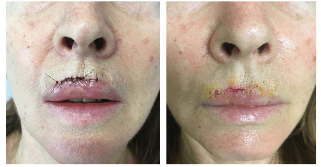
Figures 3: In post-operative period patient showed only slight oedema without neither pain nor hematoma (picture on the left), that disappeared in a few days. Cosmetic results after suture removal is shown on the right picture.

Figures 4: Aesthetic result thirty days after surgical operation.
The double advancement cutaneous-mucosal flap of our case can be considered a peculiar way of gull-wing flap, because in this case the center of the gull is based under the vermillion, not on it, but on the labial mucosa. A similar pattern of excision and arc of advancement was performed on the controlateral lip. Following excision and minor undermining, the flap was advanced and sutured superiorly, thus recreating the natural contour of the labial mucosa.
The gull-wing flap is not suitable for all upper lip defects involving Cupid’s bow. It is known that purely cutaneous defects are approached in other ways [9]. We chose gull-wing flap because our case had the two characteristics mentioned by Paniker and Mellette as inclusion criteria: vertical diameter smaller than the horizontal diameter (less than one third of the philtrum length) and location on the Cupid’s bow or near it.
Even though in literature are described many solutions to repare upper lip defects, we presented this modified gull-wing flap for its easy execution and also for the optimal cosmetic result in a cutaneous area involved in aestethic and functional purposes.
Conflicts of Interest
None.
Article Info
Article Type
Case ReportPublication history
Received: Mon 30, Mar 2020Accepted: Thu 09, Apr 2020
Published: Fri 17, Apr 2020
Copyright
© 2023 Roberto D'Astolto. This is an open-access article distributed under the terms of the Creative Commons Attribution License, which permits unrestricted use, distribution, and reproduction in any medium, provided the original author and source are credited. Hosting by Science Repository.DOI: 10.31487/j.SCR.2020.04.03
Figures & Tables
References
- Luce EA, Jing XL, Carlson T (2020) Abbe Flap Reconstruction of the Upper Lip. Plast Reconstr Surg 145: 606e-607e. [Crossref]
- Spinelli HM, Tabatabai N, Muzaffar AR, Isenberg JS (2006) Upper lip reconstruction with the alar crescent flap: A new approach. J Oral Maxillofac Surg 64: 1566-1570. [Crossref]
- Kaufman AJ, Grekin RC (1996) Repair of central upper lip (philtral) surgical defects with Island pedicle flaps. Dermatol Surg 22: 1003-1007. [Crossref]
- Kwiek B, Schwartz RA (2016) Keratoacanthoma: An Update and Review. J Am Acad Dermatol 74: 1220-1233. [Crossref]
- Paniker PU, Mellette JR (2003) A simple technique for repair of Cupid's bow. Dermatol Surg 29: 636-640. [Crossref]
- Kimble FW, Grundlingh TD (1994) A modified subcutaneously pedicled nasolabial flap for circumferential lining of the nasal vestibule. Br J Plast Surg 47: 447-449. [Crossref]
- Tian X, Xiao J, Li T, Chen W, Lin Q et al. (2017) Single-stage separation of 3- and 4-finger incomplete simple syndactyly with contiguous gull wing flaps: a technique to minimize or avoid skin grafting. J Hand Surg Am 42: 257-264. [Crossref]
- Wong RH, Agazzi S, van Loveren H (2016) “Inverted gull wing” dural closure and middle fossa floor reconstruction after transzygomatic infratemporal fossa approach. World Neurosurg 89: 280-284. [Crossref]
- Kantor J Dermatologic Surgery. McGraw Hill Education 2018: 1185-1186.