Rupture of a Rare Celiomesenteric Trunk Aneurysm following Mechanical Fall
A B S T R A C T
A celiomesenteric trunk (CMT) is an anatomical variation involving a common origin of the celiac trunk (CT) and the superior mesenteric artery (SMA). The prevalence of a celiomesenteric trunk anatomic variation has been found to be in the region of 3.4% but the incidence of aneurysms in this particular visceral artery are unknown [1]. There are only 20 cases of a celiomesenteric anomaly with an associated aneurysm documented in the last 35 years [2].
This article describes the emergency management of such an aneurysm in a frail 65-year-old female who knew about her aneurysm and was considering a recommendation for elective repair. On this occasion, the aneurysm ruptured (see figure 1) after a mechanical fall down some stairs at home. This difficult case was successfully managed with open repair and a 6mm Dacron interposition graft was sutured end-to-end with continuous 6/0 prolene from the SMA-CT common origin to the bifurcation into Splenic Artery and Common Hepatic Artery. She was discharged on post-operative day four. At her two month follow up appointment she had made an impressive recovery with no complications.
Keywords
Celiomesenteric, aneurysm, rupture, splanchnic, surgery
Case Report
A celiomesenteric trunk (CMT) is an anatomical variation involving a common origin of the celiac trunk (CT) and the superior mesenteric artery (SMA). The prevalence of a celiomesenteric trunk anatomic variation has been found to be in the region of 3.4% but the true incidence of aneurysms in this particular visceral artery are unknown [1]. There are only 20 cases of a celiomesenteric anomaly with an associated aneurysm documented in the last 35 years [2, 3].
This case report describes the emergency management of such an aneurysm in a frail 65-year-old female who knew about her asymptomatic aneurysm and was considering a recommendation for elective repair. Unfortunately, the 3.5cm diameter aneurysm ruptured (see Figure 1) after a mechanical fall down some stairs. This case was successfully managed with midline laparotomy, systemic heparinisation, and establishment of proximal and distal control with vesseloops. A 6mm Dacron interposition graft was sutured end-to-end with continuous 6/0 prolene from the SMA-CT common origin to the first large bifurcation into Splenic Artery and Common Hepatic Artery. She was discharged on post-operative day four. At six months follow up she had made an impressive recovery with no complications and had a satisfactory post-operative CT-Angiogram showing a patent interposition graft with no signs of neointimal hyperplasia or other aneurysmal change.
Discussion
This case highlights two main considerations of practice in vascular surgery: the immense variation of managing visceral artery aneurysms including in an emergency setting, and a mechanism of aneurysm rupture not commonly seen, that of mechanical fall in a frail middle-aged female. Interposition grafting using PTFE was successful in this case and the patient has had no complications or reinterventions post-operatively thus far.
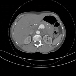
Figure 1: The celiomesenteric trunk demonstrates extraluminal extension of contrast at its left posterolateral aspect. There is significant haemorrhage adjacent to the aneurysm in the upper abdominal retroperitoneum and mesentery.
Conflicts of Interest
None.
Consent
Informed consent for this case report and associated images has been obtained from the patient.
Funding
None.
Article Info
Article Type
Case ReportPublication history
Received: Wed 08, Jan 2020Accepted: Wed 22, Jan 2020
Published: Thu 30, Jan 2020
Copyright
© 2023 James Elliott. This is an open-access article distributed under the terms of the Creative Commons Attribution License, which permits unrestricted use, distribution, and reproduction in any medium, provided the original author and source are credited. Hosting by Science Repository.DOI: 10.31487/j.CRSS.2020.01.03
Figures & Tables
References
- Wang Y, Cheng C, Wang L, Li R, Chen JH et al. (2014) Anatomical variations in the origins of the celiac axis and the superior mesenteric artery: MDCT angiographic findings and their probable embryological mechanisms. Eur Radiol 24: 1777-1784. [Crossref]
- Lipari G, Cappellari TF, Giovannini F, Pancheri O, Piovesan R et al. (2015) Treatment of an aneurysm of the celiac artery arising from a celiomesenteric trunk. Report of a case. Int J Surg Case Rep 8C: 45-48. [Crossref]
- Ailawadi G, Cowles RA, Stanley JC, Eliason JL, Williams DM et al. (2004) Common celiacomesenteric trunk: aneurysmal and occlusive disease. J Vasc Surg 40: 1040-1403. [Crossref]