Journals
Optimization of Mammary Tissue Displacement in Ultrasound Elastography
A B S T R A C T
The displacement of mammary tissues in static ultrasound elastography is often contaminated by the speckle noise deteriorating its quality. Several techniques have been developed in this context, in order to treat the noise present in images of breast tissue displacement, the progress of research work in noise processing is always questioned, especially that must be taken into account the trade-off between noise reduction and preservation of breast tissue texture. In this paper, a new strategy has been proposed to reduce speckle noise. The proposed method not only filters the image against noise, but also preserves the details and contours of the tissue texture. The approach developed is based on the coupling between the image reconstructions by filtered back projection (RPF) with an adaptive filter. The proposed model proposed has been validated on an in-vivo database comprising 20 images of the breast tissues displacement. Qualitative and quantitative improvements were noted. By comparing the proposed method with the wavelet technique, we show that it is more efficient in terms of calculating the standard deviation between the pixels (SD), it is better in terms of calculation of the Contrast / Noise ratio (CNR). And is much faster than the wavelet technique. The results of the proposed model are encouraging, and the chosen method is ready to be used in the improvement of images of mammary tissue displacements in ultrasound elastography.
Keywords
Noise of the speckle,analytical reconstruction,ultrasound elastography
Introduction
The improvement of the displacements tissues in static breast ultrasound elastography is considered as a crucial technique, facilitating the visualization of the mammary structures at the time of the pathologies research. However, elastographic imaging suffers from some limitations as to the presence of speckle noise associated with the ultrasound image [1]. Given its peculiarity in deteriorating image quality and its limitation in the visualization of small details, speckle noise is classified as one of the most complex problems in clinical diagnosis [2]. The solution is to propose a robust and efficient treatment technique to obtain a better clinical analysis of the mammary structure and tissue displacements [3]. In this context, the process of image reconstruction and image filtering of breast tissue displacements can be considered as a means of robust improvement with a good ability to eliminate noise while preserving tissue contours. The RPF of the structure of the tissue displacement of the breast followed by the adaptive filter constitutes the strategy proposed in this paper. The proposed method was validated on 20 images with tissue displacements of the breast.
The calculation of the quantitative parameters of the image such as the CNR, the SD and the execution time, shows that these parameters are better than those obtained by the wavelet technique. The proposed technique can be exploited intensively in ultrasound imaging whose purpose is to improve the diagnosis in mammary ultrasound imaging. The next section presents our reconstruction process coupled to the Wiener filter. The third section shows the results of improvement of images of tissue displacements of the breast by the proposed model, we also evaluated the quality of the images obtained qualitatively and quantitatively, by comparing them to the wavelet technique. In the fourth part, we analyzed and discussed the obtained results.
Materials & Methods
I Reconstruction by the RPF
RPF technique is used to better retrace the object and ensure that the non-visible parts are conjugated during the reconstruction, to obtain a good visibility of image details [4]. The image of tissue displacements translating the movement of the mammary tissues between less displaced rigid tissues and more displaced soft tissues, is affected by speckle noise, covering the contours of the tissues and the structures containing the diagnostic information [5]. In this perspective the application of the analytical reconstruction can solve this problem by backprojection of the elements on the same image. The reconstruction is given by the following formula: R[f(x,y)]=p(u,θ)=∫_0^Π▒〖f(x,y)dv〗 (1) Such that f (x, y) is the image of the tissue displacements, placed in the plane (x, y). "P" is the projections acquired. The axis "v" is perpendicular to the axis "u" both constituting the summation place of all the projections encountered in the image of the tissue displacements. θ is the projection angle. According to the bibliographic research, the spreading of the structures contained in the image at the time of the overhead projection, produces spreading residues. To manage this limit, the use of a ramp filter can eliminate these residues [6]. RPF allows reconstructing structures in the image to ensure better visualization of lesions. However the noise is still present in the image which led us to seek a filtering technique.
II Adaptive filtering
The presence of speckle noise in the image obscures the small difference in gray level and degrades its quality. The properties of the speckle noise had been discussed by the researchers with a view to finding a conclusive solution. It is a multiplicative noise that constitutes a significant limit in clinical diagnosis. Therefore, filtering becomes an important step before proceeding to the analysis of mammary pathologies. We have proposed the implementation of the Wiener filter following a reconstruction by RPF, it is a linear filter, which applies its Wiener function in an adaptive way, when the local image variance is important, the filter performs little smoothing, when the local image variance is small, the filter performs more smoothing [7]. The proposed adaptive filter is more selective than a comparable linear filter. Its application reduces noise while preserving the edges of breast tissue [8]. The Wiener window estimates the mean and the local variance in each pixel according to the following formula: μ=1/NM ∑_(n1,n2)▒〖a(n1,n2)〗 (2) σ²=1/NMΣn1n2a²(n1,n2)-µ² (3) Such that N and M respectively represent the number of rows and the number of columns defining the image matrix. The Wiener filter window is defined by the estimation formula below: b(n1,n2)=µ+σ²-ν²/σ²(a(n1,n2)-µ) (4) Such as V2 is the variance of speckle noise, and if noise variance is not available, the Wiener filter window uses the range of all estimated variance.
Results
The proposed method was applied to 20 images of breast tissue displacements with malignant tumors, these results were validated by two radiologists.
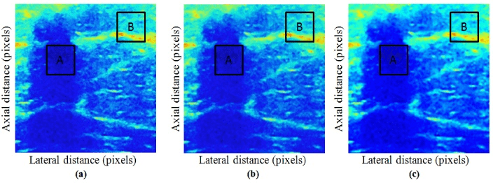
Figure 1: Effect of image enhancement methods of breast tissue displacements. a Image of original untreated tissue displacements. b Image of tissue displacements obtained by wavelet technique. c Image of tissue displacements obtained by the proposed method.
The adopted and proposed method in this paper was analyzed and evaluated using quantitative criteria. In order to confirm the occurrence and efficacy of the proposed technique, we compared it to a method of processing wavelet speckle noise: a method used in image processing of tissue displacements. Quantitative measures include EA, CNR and execution time.
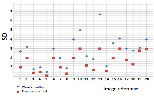
Figure 2: Results of calculation of the 20-image SD showing tissue displacements for both methods: wavelet method and proposed method.
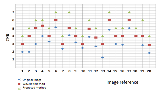
Figure 3: CNR calculation results of 20 images showing tissue displacements for both methods: wavelet method and proposed method.
Discussion
In this section we will compare and discuss the obtained results. According to (Figure 1), the displacement mammary tissues shows a strong disappearance of speckle noise in the case of the proposed method, showing a structure devoid of artifact, with good preservation of the contours. The proposed method was evaluated by analyzing the quantitative criteria of the images in comparison with the results of the wavelet method. From (Figure 2), the SD between the pixels of the wavelet method is greater than that obtained by the proposed method. This is explained by the perfect adaptive filtering translating the minimum error obtained in the proposed model. The adaptive filter placed after the RPF allows having more similar tissue structures to the origin with a good visualization of details, explaining the obtained result.
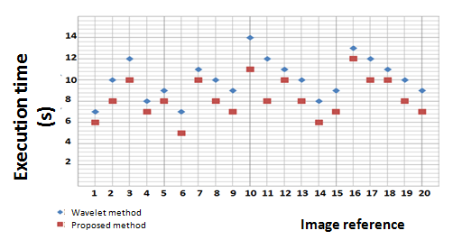
Figure 4: Results of calculation of the execution time of the two methods: wavelet method and proposed method, for 20 images showing tissue displacements.
From (Figure 3), the CNR of the proposed method is superior to those obtained in the original image and in the wavelet technique. This result is highly anticipated since the RPF ensures the improvement of the contrast by reconstruction of the objects in the image. For each stack obtained, a new tracing of the texture is added, which suddenly improves the contrast. Added to this, the adaptive filter which allows reducing the noise in a very important way, which also contributes to increase the CNR proportionally. This improvement no longer exists in the wavelet processing technique, since this method reduces noise without preserving the contrast of low echogenicity borders.
The last quantitative criterion corresponds to the execution time required for each method. The simulation results were performed on a MATLAB, run on a Pentium 4 computer, 4.2 GHz, 6 GB RAM on Windows 7. From (Figure 4), the analysis of the speed of each method in terms of calculation the speed of execution, shows that the method proposed is faster than the method using the wavelets. The wavelet technique decomposes the image according to a selected threshold, this requires an expensive calculation time in order to completely filter the image. On the other hand, the proposed method reconstructs the structures of the image, and the filter directly by adaptive filtering, which quickly reduces the noise without falling into a slow calculation time. The proposed method has been validated qualitatively by two radiologist doctors, and quantitatively by the calculation of the criteria mentioned above. The proposed model will produce good quality images with minimal error between pixels, better CNR and faster execution time than wavelet technique.
Conclusion
In this article, a new model for improving the quality of the breast tissue displacement image has been proposed. The adopted strategy is to couple a reconstruction by RPF with an adaptive filter. We have shown that the proposed method can effectively reduce speckle noise, the results were validated using 20 images showing breast tissue displacements. Quantitative criteria (SD, CNR and execution time) showed greater performance superiority over the wavelet technique. The proposed approach can be considered as a good tool in image processing of breast tissue displacements, the implementation of this method will help to ensure accurate diagnosis, leading to create an adequate platform in ultrasound elastography of the breast. As a perspective of this work, we propose to improve much more the quantitative parameters of the image, by the development of the techniques of filtering and improvement of the reconstruction of the mammary structure, using iterative algorithms with a computation time short.
List of abbreviations
RPF: Filtered Back Projection SD: Standard Deviation between the pixels CNR: Contrast, Noise ratio
Ethics approval and consent to participate
The radiology department of the Hospital University Monji Slim Marsa and the Laboratory of Biophysics and Medical Technology Research declare agreement on ethical approval and consent: 20 images without the names of patients (anonymous) contain malignant tumors in the breast. The ethics committee of the laboratory of Biophysics and Medical Technologies, reference number LR13ES07 has approved the study. Written informed report was obtained from the patients for publication of this manuscript and accompanying images.
Consent for publication
Not applicable.
Availability of data and material
All data analysed during this study are included in this published article.
Competing interests
The authors declare that they have no competing interests.
Funding
This research did not receive any specific grant from funding agencies in the public, commercial, or not-for-profit sectors.
Acknowledgements
The authors would like to thank the doctors teams of the university hospital of Monji Slim,Tunisia, for providing and assessing the clinical Breast data.
Authors' Information
Professor Taher Slimi is a professor in medical imaging also an engineer in medical imaging and a Biophysicist. Professor Romaissa Tamali is a professor in medical imaging and breast cancer treatment Professor Halima Mahjoubi is a Professor in Biophysics and Medical Imaging.
Article Info
Article Type
Research ArticlePublication history
Received: Thu 16, May 2019Accepted: Wed 03, Jul 2019
Published: Fri 19, Jul 2019
Copyright
© 2023 Taher Slimi. This is an open-access article distributed under the terms of the Creative Commons Attribution License, which permits unrestricted use, distribution, and reproduction in any medium, provided the original author and source are credited. Hosting by Science Repository.DOI: 10.31487/j.RDI.2019.03.07
Author Info
Corresponding Author
Taher SlimiLaboratory of Techniques and Medical Complexity, Faculty of Medicine, Joseph Fourier University, Grenoble, France
Figures & Tables
References
- Eisenbrey JR, Dave JK, Forsberg F (2016) Forsberg F.Recent technological advancements in breast ultrasound. Ultrasonics 70: 183-190. [Crossref]
- Slimi, Marzouk, Kraiem, Mahjoubi (2017) BioMed Eng OnLine 16: 19.
- Alessandrini M, Bernard O, Basarab A (2013) Multiscale optical computation from the monogenic signal. AEMB 34: 33-37.
- N Chetih, Z Messali (2015) Tomographic image reconstruction using filtered back projection (FBP) and algebraic reconstruction technique (ART). Int Conference Control 1-6.
- Kuo Y, Lin YY, Lee RC, Lin CJ, Chiou YY et al. (2016) Comparison of image quality from filtered back projection, statistical iterative reconstruction, and model-based iterative reconstruction algorithms in abdominal computed tomography. Medicine (Baltimore) 95: e4456 [Crossref]
- M. Pérez-Liva, JL. Herraiz, L. Medina-Valdés (2015) Regularization of image reconstruction in ultrasound computed tomography. Nuclear Sci Symposium Med Imagin Confer 1-3.
- Slimi, Marzouk, Kraiem, Mahjoubi (2017) Estimation of Displacement Tissues in Breast Ultrasound Ultrasound Elastography. J Diagn Tech Biomed Anal 6: 1.
- J. Clerk Maxwell (1892) A Treatise on Electricity and Magnetism. 68-73.