Operative Removal of 154 Objects in Chronic Compulsive Foreign Body Ingestion
A B S T R A C T
Intentional foreign body ingestion presents as a chronic surgical challenge. We encountered a severe case of ingestion that had failed endoscopic retrieval secondary to the size, shape, and quantity of structures encountered. Open gastrostomy with removal of objects with care to prevent future hostile abdomen was performed with 154 total objects removed at completion. We report a lesson in approaching a clinical problem with thought to approach for the preservation of anatomical structures in potential future interventions.
Keywords
Foreign body ingestion, open gastrotomy
Introduction
Eighty percent of cases of foreign body ingestion are seen in children, with the majority of these ingestions being accidental [1, 2]. Cases of intentional foreign body ingestion are less common but are most frequently seen amongst the adult population. Many of these patients have psychiatric disorders, a history of substance use, developmental delay, or are incarcerated [1, 3]. Studies have shown that up to 85% of patients presenting with intentional ingestion had an underlying psychiatric condition [4]. Many of this patient population also have a history of prior intentional ingestion, and often present with multiple ingested objects on arrival [1]. Amongst reported cases, an assortment of objects has been intentionally ingested by patients including pens, pencils, utensils (spoons, forks, knives), toothbrushes, batteries, razor blades, coins, hardware (screws and bolts), pieces of glass, and paper clips [2-4].
Many factors must be taken into consideration when devising a management plan for intentional foreign body ingestion. These include patient age, symptoms, type of foreign body ingested, location of the foreign body within the GI tract, amount of time passed since ingestion, and associated complications such as hemorrhage, bowel obstruction, and GI perforation [2, 4]. At initial presentation, patients may complain of epigastric pain, vomiting, dysphagia, chest pain, or may be asymptomatic [2]. Objects are most commonly found within the esophagus, stomach, or duodenum [2-4].
Depending on the specific case, the patient may be managed conservatively, endoscopically or surgically. Reports published previously have largely focused on accidental foreign body ingestions; studies show that 80-90% of ingested foreign bodies pass spontaneously, 10-20% require endoscopic intervention, and about 1% of cases are managed surgically [5]. Recent studies show that the rate of surgical intervention in the setting of intentional ingestion ranges from 12-16%, which is higher than in cases of accidental ingestions. This may be due to the choice of ingesting more dangerous objects in intentional cases to inflict self-harm [2]. In one study of 262 cases of intentional foreign body ingestion at an urban county hospital, 14% were managed conservatively, 76% underwent endoscopy, 7% required surgical intervention upon initial presentation for complications such as perforation, and 3% received unknown management. An additional 4% of patients who initially underwent unsuccessful endoscopic management went on to receive surgical management, totaling 11% of cases that were managed surgically [4].
Case Presentation
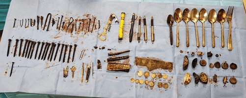
A 25-year-old male with no known past medical or surgical history presented from home with a two-week history of nausea, vomiting, and decreased oral intake. He complained of abdominal pain but refused to answer any other questions. Family members stated he had a history of drug use and alcohol abuse but had moved in with family and not used drugs or alcohol in three months. He had never had surgery, taken medications, or been diagnosed with developmental or abnormal behaviours. On exam, his abdomen was mildly tender and distended, but otherwise appropriate. An abdominal radiograph was obtained that showed a distended stomach with multiple radiopaque objects concerning for foreign objects. Gastroenterology was consulted who performed an emergent endoscopy. Multiple objects were able to be identified including several pieces of silverware, bottle caps, a wine opener, and a phone charger, but only the phone charger was able to be safely extricated without injury to the esophagus. Acute Care Surgery was consulted for operative intervention. He was taken to the OR and positioned with a bump under his left side. A small supraumbilical midline laparotomy was made and the stomach was able to be identified and brought midline with two laps positioned posteriorly. Two 2-0 silk sutures were placed anteriorly approximately 4 cm apart and using electrocautery, a small gastrotomy was made. Using ring forceps, one hundred and fifty-four objects were removed (Figure 1). The stomach lining was examined with multiple areas of thickened scarring and mild gastritis. A C-arm was used for intra-operative imaging to determine that all objects had been completely removed. The gastrotomy was closed in two layers. Prior to closing the abdomen, an endoscopy was used to perform a complete exam of the stomach and duodenum, and a completion leak test.
Figure 1: Gastrotomy contents retrieved.
Finally, a nasogastric tube was placed and secured in place. The laparotomy incision was closed, and the patient was extubated. Post operatively the patient received an IV proton pump inhibitor twice daily and remained NPO with nasogastric tube to low intermittent wall suction for five days. On day five, he was given a clear liquid diet and advanced as tolerated. A psychiatric consult was obtained, and unfortunately there is no medical or cognitive therapy to prevent or deter this behaviour at this time. He was discharged to home with family.
He was healing well at his two weeks follow up and continues to deny swallowing objects however one-month post op, re-presented after swallowing a crescent wrench which was able to be removed endoscopically.
Discussion
Patients who compulsively swallow inanimate objects present a unique surgical patient population that requires forethought with planning. Without medical or cognitive therapy, this disease is often chronic, and will require repeat operations. Incision planning to prevent adhesion formation and limit breakdown of the abdominal wall and gastric wall integrity are imperative. Intraoperatively, a leak test to rule out micro-perforation from sharp objects and removal limits re-operation and missed injury. Post operatively, due to the mechanical trauma of the foreign objects on the gastric mucosa, prolonged decompression and limiting acid production with prescribed PPI at discharge allow for a well healed gastrotomy and ongoing preventative efforts in a chronic surgical patient. While this patient intra-operatively may be straight forward, it is the understanding and planning for potential future operations that makes this patient a unique acute care surgical patient that deserves our forethought upon clinical exam.
Article Info
Article Type
Case ReportPublication history
Received: Mon 10, Aug 2020Accepted: Mon 21, Sep 2020
Published: Fri 02, Oct 2020
Copyright
© 2023 Amanda Chelednik. This is an open-access article distributed under the terms of the Creative Commons Attribution License, which permits unrestricted use, distribution, and reproduction in any medium, provided the original author and source are credited. Hosting by Science Repository.DOI: 10.31487/j.AJSCR.2020.04.01
Author Info
Zahra Haider Amanda Chelednik Jacob Quick
Corresponding Author
Amanda ChelednikDepartment of Acute Care Surgery, University of Missouri-Columbia, Columbia, Missouri, USA
Figures & Tables

References
- Palese C, Al Kawas FH (2012) Repeat intentional foreign body ingestion: the importance of a multidisciplinary approach. Gastroenterol Hepatol (N Y) 8: 485-486. [Crossref]
- Dalal PP, Otey AJ, McGonagle EA, Whitmill ML, Levine EJ et al. (2013) Intentional foreign object ingestions: need for endoscopy and surgery. J Surg Res 184: 145-149. [Crossref]
- Huang BL, Rich HG, Simundson SE, Dhingana MK, Harrington C et al. (2010) Intentional swallowing of foreign bodies is a recurrent and costly problem that rarely causes endoscopy complications. Clin Gastroenterol Hepatol 8: 941-946. [Crossref]
- Palta R, Sahota A, Bemarki A, Salama P, Simpson N et al. (2009) Foreign-body ingestion: characteristics and outcomes in a lower socioeconomic population with predominantly intentional ingestion. Gastrointest Endosc 69: 426-433. [Crossref]
- Webb WA (1988) Management of foreign bodies of the upper gastrointestinal tract. Gastroenterology 94: 204-216. [Crossref]
