Journals
An Unusual Case of Humeral Shaft Non-Union
A B S T R A C T
Background: Humeral shaft fractures account for 1% to 3% of all fractures and approximately 20% of all fracture involving the humerus. The prevalence of non-union for diaphyseal humeral fractures has been reported as 1% to 10% after non-surgical and 10% to 15% after surgical management. Various devices used in treatment of humeral diaphyseal non-union are limited contact dynamic compression plates, locking compression plate (LCP), wave plates, humerus interlocking nail (IMN), Ilizarov external fixators and bone graft struts.
Case Description: A 68-year-old man reported a humeral shaft fracture on the left side, due to a simple fall. It was reduced and fixed by IMN. He underwent clinical and radiological follow up. Three months after the intervention, due to persistent pain and impaired function of the left shoulder, the nail was removed and a cemented endoprosthesis was implanted. 3 years later, unsatisfied with the results, he came to our attention and was diagnosed an atrophic non-union in the site of the previous humeral shaft fracture. Moreover, the exams showed a rotator cuff insufficiency. It was decided to perform a single-stage intervention to achieve two goals: cure the humeral shaft non-union and restore the function of the left shoulder. The cemented endoprosthesis was removed, followed by an extensive curettage of the non-union site. A reverse prosthesis was implanted, with an extra-long stem used to stabilize the non-union site, as it was an IMN. An allograft was harvested from a cadaver femur and fixed with two metal cerclages. The patient underwent clinical and radiological follow-up. Complete healing was achieved 8 months later.
Conclusion: Humeral shaft nonunion still represent a pathology that pose a serious problem to the surgeon. A correct management should include an accurate pre-operative planning, to achieve the best result possible for the patient.
Keywords
Humeral shaft, fracture, intramedullary nail, non-union, allograft, reverse total shoulder prosthesis
Introduction
Humeral shaft fractures account for 1% to 3% of all fractures and approximately 20% of all fracture involving the humerus [1, 2]. The choice for the optimal treatment is still controversial. Non operative treatments include functional bracing, casting, and splinting. Operative treatments currently viable are plate fixation (open reduction internal fixation ORIF), intramedullary nailing (IMN) and external fixation [3]. Among these, no gold standard treatment has been identified yet, and non-union may occurs. The prevalence of nonunion for diaphyseal humeral fractures has been reported as 1% to 10% after non-surgical and 10% to 15% after surgical management [4].
Causes of humeral diaphyseal fracture non-unions are infection, distraction at fracture site, soft tissue interposition, unstable fixation, wrong choice of implant, iatrogenic devitalization of soft tissues, inadequate immobilization, open fractures, comminution, and osteoporosis [5]. Various devices used in treatment of humeral diaphyseal non-union are limited contact dynamic compression plates, locking compression plate (LCP), wave plates, humerus interlocking nail (HIL), Ilizarov external fixators and bone graft struts. Currently, the most popular management for a humeral shaft non-union is ORIF with autologous bone grafting [6]. In this paper we present an unusual case of humeral diaphyseal fracture non-union.
Case Description
A 68-year-old male, unemployed, on July 2014, reported a spiroid fracture of the left humeral diaphysis (AO/Muller classification: 12-C1) due to a simple fall from a ladder. Some days later he underwent surgery, and the fracture was fixed by IMN (Figure 1). Three months later, he was clinically and radiologically followed up (Figure 2). The patient reported pain, discomfort, and function impairing, but he was suggested to continue the rehabilitation program. 7 months later, due to the increasing pain and functional impairing, the patient was diagnosed a post-traumatic humeral head avascular necrosis with an intact rotator cuff. For this reason, the IMN was removed and a cemented shoulder endoprosthesis was implanted.

Figure 1: IMN of the humeral shaft fracture, post-operative radiographic check.

Figure 2: 3 months X-ray follow up, showing lack of consolidation in the site of the fracture.
All these information above reported were obtained directly from the patient and from his clinical records. For this reason, it was not possible to gain access to all the imaging studies performed. On April 2017, the patient came to our attention, complaining persistent impaired function on the left shoulder despite the prosthesis. The clinical and radiological evaluation was impressive (Figure 3). The patient discomfort was due to the presence of a “second elbow” in the middle of the arm with a preternatural mobility caused by an atrophic non-union of the humeral shaft. On July 2017, 3 years after the first operation, the patient underwent new surgery.

Figure 3A: clinical examination showing a multiplanar preternatural mobility in the middle of the arm.

Figure 3B: X rays showing the nonuion in the site of the previous fracture. The head of the endoprosthesis has migrated upward, a sign meangingfull of rotator cuff insufficiency.

Figure 3C: CT-scan with 3D reconstruction highlightning an atrophic nonunion of the humeral shaft, with a significant bone gap.
The main goals of this reintervention were to treat the humeral shaft non-union and restore the shoulder function. It was decided to gain these aims in only one stage. After removing the previous prosthesis and its cement, fracture intramedullary fixation was obtained with the extra-long stem (180 mm) of the reverse shoulder arthroplasty (SMR model, LIMA corporate, Italy) we implanted to restore shoulder function. The extra-long stem was chosen to include and surpass the non-union site, which was bonified with an extensive curettage. Due to the relevant bone loss, an allograft from a cadaver femur, was prepared and fixed with two metal cerclages (Figure 4). The patient was dismissed with the upper arm immobilized by a sling for six weeks. Active kinesis for the elbow and the wrist were allowed. Passive kinesis of the shoulder started after six weeks. The patient underwent radiological and clinical examination every month (Figure 5), and 8 months after the intervention radiographical healing was obtained.

Figure 4A: Intra-operative image showing the previous implanted endoprosthesis, with the atrophic nonunion at the lower extremity. Notice the wide bone gap in the middle of the humers. An extended delto-pectoral approach was used.

Figure 4B: In the square, preparation of the bone splint from the cadaveric femur. Intra-operative image showing the positining of the allograft (A) and its fixation by metal cerclages (arrows). Notice the head of the reverse prosthesis (P), implanted before the fixation of the allograft.
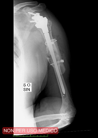
Figure 5A: Post-operative check, showing the correct positioning of the reverse prosthesis and the allograft.

Figure 5B: 3 months radiological follow up, showing initial sign of consolidation.
Discussion
The most relevant findings in this report are the use of a revision stem with an allograft to treat a humeral shaft non-union and the importance of a correct pre-operative planning. In this case there were two goals to achieve: firstly, to cure the humeral shaft non-union, secondly to restore the function of the shoulder. These goals could have been achieved in two different ways: the first was a two or even three stages process with multiple surgical interventions, the second was a one-stage way with a single intervention able to solve both the problems at the same time. Crucial in the decision making was how to treat the nonunion. Currently, the most popular management for a humeral shaft non-union is ORIF with autologous bone grafting, even if no consensus has been reached regarding the best treatment [6, 7]. IMN has also been used. Martinez et al., comparing the locking plate vs IMN, achieved 100% union rate and had minimal complications with both methods, and reported that nailing achieved earlier union with fewer complications [8]. Singh et al. also achieved comparable functional outcome between ORIF and IMN [6].
Another crucial point in the decision making, was the choice of the graft to use. When positioning the plate, an autograft is usually used, generally from the iliac crest. In the elderly, where the poor bone quality could represent a contraindication for harvesting the iliac crest, the use of allograft was recently advised [7]. The decision about the best way to treat nonunion was influenced by the second goal to achieve, which was to restore the function of the impaired shoulder. To do this, it was necessary to remove the cemented endoprosthesis and implant a new one. Due to the rotator cuff insufficiency, a reverse prosthesis was chosen.
As it was mentioned before, there were two ways to achieve nonunion healing and restore the function of the shoulder. Choosing the plate as method of treatment would have meant performing at least two to three surgical procedure: the first one to remove the endoprosthesis, treat the nonunion and implanting the plate, an eventually second one to remove the plate and finally a third procedure to implant the new reverse prosthesis. It’s easy to imagine the amount of physical stress this way would cause to the patient, and the amount of time needed to perform it.
In this case it was decided to perform a single-stage procedure, which allowed us to save a significant amount of time and prevent the patient to undergo several interventions, also reducing the infective risk. The choice of a revision stem for the reverse prosthesis was made to use it as an IMN. As above illustrated, the extra log stem allowed us to surpass the nonunion site to give stability to the humerus. Due to the extensive curettage performed, an autograft from the iliac crest was not feasible. It was decided to use a cadaveric femur to harvest an allograft, which was carefully shaped to obtain a splint-like shape. This splint was then fixated to the humerus in a “plate-fashion” way, so to give more stability to the construct. The choice of using metallic cerclages to fix it, was made to preserve bone stock from an already poor site.
A limitation of this case report was the impossibility to fully reconstruct the patient history before April 2017, due to lack of radiological images and clinical information. For these reasons it was difficult to analyze properly the decision-making process which guided the initial surgery.
Conclusion
Humeral shaft nonunion still represent a pathology that pose a serious problem to the surgeon. Today no consensus has been reached on the best way to treat them, although most surgeons seems to prefer ORIF with autologous bone graft. A correct management should include an accurate pre-operative planning, to achieve the best result possible for the patient.
Conflicts of Interest
None.
Funding
None.
Ethical Approval
All procedures performed in studies involving human participants were in accordance with the ethical standards of the institutional and/or national research committee and with the 1964 Helsinki Declaration and its later amendments or comparable ethical standards.
Consent
Patient gave his informed consent.
Article Info
Article Type
Case ReportPublication history
Received: Thu 25, Jun 2020Accepted: Fri 10, Jul 2020
Published: Thu 23, Jul 2020
Copyright
© 2023 Claudio Chillemi. This is an open-access article distributed under the terms of the Creative Commons Attribution License, which permits unrestricted use, distribution, and reproduction in any medium, provided the original author and source are credited. Hosting by Science Repository.DOI: 10.31487/j.IJSCR.2020.03.02
Author Info
Corresponding Author
Claudio ChillemiDepartment of Orthopaedic Surgery, Istituto Chirurgico Ortopedico Traumatologico (ICOT), Latina, Italy
Figures & Tables
References
- Ward EF, Savoie FH, Hughes JL (1998) Fractures of the diaphyseal humerus. In: Skeletal trauma: fractures, dislocation, ligamentous injuries. Philadelphia: Saunders 2: 1523-1547.
- S H Rose, L J Melton 3rd, B F Morrey, D M Ilstrup, B L Riggs (1982) Epidemiologic features of humeral fractures. Clin Orthop Relat Res 168: 24-30. [Crossref]
- T Apivatthakakul, O Arpornchayanon, S Bavornratanavech (2005) Minimally invasive plate osteosynthesis (MIPO) of the humeral shaft fracture. Is it possible? A cadaveric study and preliminary report. Injury 36: 530-538. [Crossref]
- Jeffrey Richmond, Kevin Colleran, Olivier Borens, Peter Kloen, David L Helfet (2004) Nonunions of the distal tibia treated by reamed intramedullary nailing. J Orthop Trauma 18: 603-610. [Crossref]
- Cleveland KB (2008) Delayed union and non union of fractures. In: Canale ST, Beaty JH (eds) Campbell’s operative orthopaedics, 11th edn, Philadelphia. Mosbyp 3: 3529-3565.
- Ashutosh Kumar Singh, G R Arun, Nidhi Narsaria, Anurag Srivastava (2014) Treatment of non-union of humerus diaphyseal fractures: a prospective study comparing interlocking nail and locking compression plate. Arch Orthop Trauma Surg 134: 947-953. [Crossref]
- Giuseppe Toro, Federica Lepore, Giampiero Calabrò, Gabriella Toro, Marco Rossini et al. (2019) Humeral shaft non-union in the elderly: Results with cortical graft plus stem cells. Injury 50: S75-S79. [Crossref]
- Angel A Martínez, Jorge Cuenca, Antonio Herrera (2004) Treatment of humeral shaft nonunions: Nailing versus plating. Arch Orthop Trauma Surg 124: 92-95. [Crossref]