A Rare Case of Urachal Remnant with Calculus Just Below Umbilicus
A B S T R A C T
Introduction: Urachal remnants, such as the vesicourachal diverticulum, or urachal cysts are rarely involved in calculus or calcification. There are currently no case reports of urachal remnants with calculus just below the umbilicus.
Case Presentation: A healthy 28-year-old man presented with umbilical drainage. Abdominal computed tomography showed inflammation just below the umbilicus with calculus. The patient was diagnosed with ombilitis with a urachal remnant and underwent laparoscopic resection for the urachal remnant after the attenuation of ombilitis. Surgical findings showed that the omentum adhered to the abdominal wall along with the urachal duct. The urachal duct with calculus was resected at the top of the bladder. The main component of calculus was silicic acid. Pathological findings were compatible with a urachal remnant and there was no evidence of a malignant tumor.
Discussion: The main component of calculus is derived from necrotic material caused by inflammation because silicic acid was detected, indicating a different etiology from that of urinary calculus.
Conclusion: We encountered a rare case of a urachal remnant with calculus below the umbilicus.
Keywords
Urachal remnant, ombilitis, calculus, calcification
Introduction
Urachal remnants are associated with repeated relapse and rarely lead to a malignant tumor [1]. Case reports of urachal remnants, such as the vesicourachal diverticulum, or urachal cysts together with calculus or calcification have been reported in the literature [2, 3]. We herein describe a rare case of a urachal remnant with calculus below the umbilicus.
Case Report
A 28-year-old man presented with umbilical drainage. Abdominal computed tomography (CT) showed localized inflammation just below the umbilicus with calculus (Figure 1). The patient was diagnosed with ombilitis due to a urachal remnant. Laparoscopic resection of the urachal remnant was performed.
I Surgical Findings
The omentum adhered to the abdominal wall along with the urachal duct (Figure 2a). The urachal duct and bilateral medial umbilical fold were identified after the detachment of adhesions (Figure 2b). The urachal duct was resected at the top of the bladder (Figure 2c) and the umbilical remnant with calculus was removed through the incision of the umbilicus.
II Resected Specimen Findings
The urachal remnants with calculi were clumped together due to inflammation (Figure 3). The main component of calculus was silicic acid.
III Pathological Findings
There was no evidence of a malignant tumor. A luminal structure compatible with a urachal remnant was identified. The patient was discharged on postoperative day 4 without any complications.
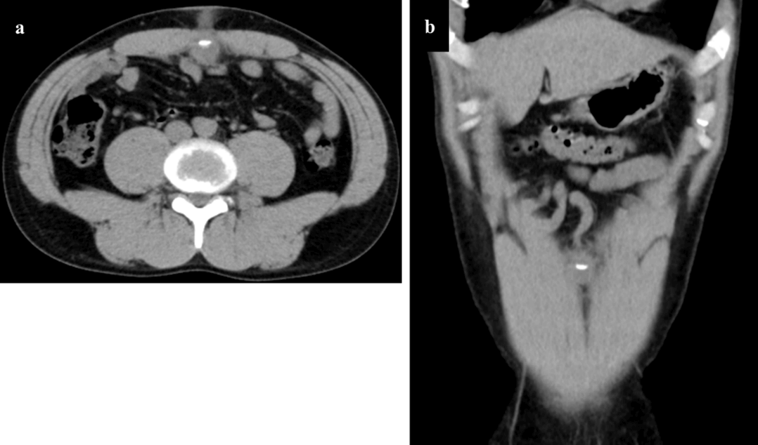
Figure 1: Abdominal CT showing mass with calculus just below the umbilicus.

Figure 2: a) The omentum adhered to the abdominal wall along with the urachal duct. b) The urachal duct (arrowheads) and bilateral medial umbilical fold (arrows) were identified. c) The urachal duct was resected at the top of the bladder (arrowheads).
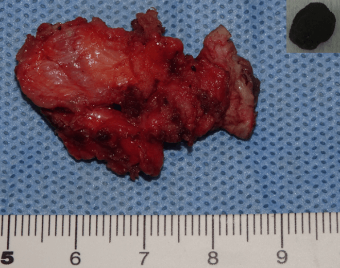
Figure 3: The urachal remnants with calculi were clumped together due to inflammation.
Discussion
The urachal duct, an epithelial luminal structure that connects the umbilicus to the bladder, is generally formed in the early stages of fetal life and regresses after approximately 8 weeks of gestation [4]. Urachal remnants are classified into four types based on their morphology: patent urachus, which is open from the bladder side to the umbilical side; urachal cyst, which is closed at both ends; umbilical-urachus sinus, which is open only at the umbilical side; and vesicourachal diverticulum, which is open only at the bladder side [5]. Patients with urachal remnants are mostly asymptomatic, but may become symptomatic with abdominal pain, redness, swelling, or drainage when complicated by infection [6]. Our patient was diagnosed with ombilitis due to umbilical-urachus sinus because abdominal CT showed localized inflammation around the umbilicus.
Urachal remnants are indicated for surgical treatment because they are often accompanied by omphalitis, which sometimes recurs when treated by drainage alone. Furthermore, urachal remnants may progress to cancer [7]. Urachal remnants with calculus are very rare, but suggest communication between urachal remnants and bladder, allowing urine to flow into the urachal lesion [2]. Moreover, calcification or calculus within urachal remnants is frequently associated with tumors [8]. In the present case, CT did not reveal any findings of a tumor, and surgery was performed for the urachal remnant.
If the main component of calculus is derived from urinary calculus, urine comes into contact with the shed epithelium filling the lumen of the urachal duct, and salts in urine precipitate and become a nucleus for the formation of calculus. However, in the present case, pathological findings did not reveal a lumen between the bladder and urachal duct. Therefore, calculus formed with necrotic material caused by inflammation because silicic acid was detected, indicating a different etiology from that of urinary calculus.
Conclusion
We encountered a rare case of a urachal remnant with calculus that formed directly below the umbilical region with necrotic material due to omphalitis caused by the urachal remnant.
Acknowledgment
Not applicable.
Conflicts of Interest
None.
Funding
None.
Ethical Approval
Not applicable.
Availability of Data and Materials
Not applicable.
Consent
Written informed consent was obtained from the patient for the publication of this case report and any accompanying images.
Guarantor
The first author is Dr Hirotaka Kato, who accepts full responsibility for the work and/or the conduct of the study.
Author Contributions
Hirotaka Kato analysed and interpreted the patient data, made figures, and wrote the manuscript. Masaaki Deguchi and Yoshifumi Sakata supervised, read and approved the final manuscript.
Article Info
Article Type
Case ReportPublication history
Received: Thu 01, Sep 2022Accepted: Tue 20, Sep 2022
Published: Tue 27, Sep 2022
Copyright
© 2023 Hirotaka Kato. This is an open-access article distributed under the terms of the Creative Commons Attribution License, which permits unrestricted use, distribution, and reproduction in any medium, provided the original author and source are credited. Hosting by Science Repository.DOI: 10.31487/j.JSCR.2022.02.03
Figures & Tables
References
1. Bruins HM, Visser
O, Ploeg M, Hulsbergen van de Kaa CA, Kiemeney LA et al. (2012) The clinical
epidemiology of urachal carcinoma: results of a large, population based study. J
Urol 188: 1102-1107. [Crossref]
2. Ozbülbül NI, Dağli
M, Akdoğan G, Olçer T (2010) CT urography of a vesicourachal diverticulum
containing calculi. Diagn Interv Radiol 16: 56-58. [Crossref]
3. Seo IY, Han DY, Oh
SJ, Rim JS (2008) Laparoscopic excision of a urachal cyst
containing large stones in an adult. Yonsei Med J 49:
869-871. [Crossref]
4. Ashley RA, Inman
BA, Routh JC, Rohlinger AL, Husmann DA et al. (2007) Urachal anomalies: a
longitudinal study of urachal remnants in children and adults. J Urol
178: 1615-1618. [Crossref]
5. Blichert Toft M,
Koch F, Nielsen OV (1973) Anatomic variants of the urachus related to clinical
appearance and surgical treatment of urachal lesions. Surg Gynecol Obstet
137: 51-54. [Crossref]
6. Milotic F, Fuckar
Z, Gazdik M, Cicvaric T, Milotic I et al. (2002) Inflamed urachal cyst
containing calculi in an adult. J Clin Ultrasound 30: 253-255. [Crossref]
7. Watanabe T, Ishigure K, Fujioka A, Horiba T, Hirai T et al. (2008) A case of urachal carcinoma presented with an abscess of a urachal remnant. J Jpn Surg Assoc 69: 175-178.
8. Rodrigues JC, Gandhi S (2013) Don't get caught out! A rare case of a calcified urachal remnant mimicking a bladder calculus. J Radiol Case Rep 7: 34-38. [Crossref]