Journals
A Giant Enchondroma Mimicking Sarcoidosis: Report of Case and Review of the Literature
A B S T R A C T
Sarcoidosis of the humerus is exceptionally rare and only a few cases have been reported. In this mini-review, a case of enchondroma in proximal humerus mimicking sarcoidosis and the features of bone involvement of sarcoidosis were reviewed.
A 41-year-old female who was diagnosed with sarcoidosis in 2009 had used corticosteroids for 4 months. She had not any symptoms until 2014. She was admitted the clinic with a 2-month-history of erythema nodosum on her legs and right shoulder pain. Values of laboratory tests were all within normal limits except erythrocyte sedimentation rate and C-reactive protein levels. The mass in the proximal metaphyseal humerus has the characteristic as a medullary lesion which had calcifications in CT sections. MR images that the mass had low signal intensity on T1-weighted images and heterogeneous high signal intensity on T2-weighted images. It had irregular nodular contrast and the mass did not cause the expansion of the bone. Increased activity was observed on scintigraphy. We suspected the mass which was realized incidentally in a patient with sarcoidosis, could be bone sarcoidosis. As a result of our biopsy, it was enchondroma.
Bone neoplasms should be kept in mind in issues like our case report.
Keywords
Bone,enchondroma,sarcoidosis
Introduction
Sarcoidosis is an autoimmune multisystemic inflammatory and granulomatous disease and. its etiology is still unclear [1]. Proposed pathogenesis includes a combination of genetic, immunologic, and environmental factors. However, environmental and immunologic factors, either infectious or non-infectious, affect individuals with genetic predispositions to cause chronic inflammatory responses, leading to the disease onset. Immunologically, locally active cellular immunity with systemically impaired cellular immunity, indicated by (-) Tuberculin test, is remarkable. Hypergammaglobulinemia is usually accompanied and anti-nuclear antibodies and rheumatoid factor are detected in significant number of patients [2]. Sarcoidosis generally affects young and middle-aged patients, and women have a higher prevalence than men. There are two peaks, one is between 20-29 years and the other is for women over age 50 years [3].
Lungs (is diseased in over 90% of patients), mediastinal lymph nodes, skin, and eyes are the mainly affected organs [3]. Furthermore, it may affect bones, joints, parotid glands, liver, and kidneys. In the other hand, any organ virtually can be affected [4]. Clinical symptoms and findings are highly divergent depending on the affected organ, including facial nodules and nodular erythemas in the skin, blurred vision and myodesopsia in eyes, headache and paralysis in the CNS, and atrioventricular block in the heart. Bilateral hilar lymphadenopathy in chest X-ray increased numbers of total cells and lymphocytes and an elevated CD4/8 ratio in bronchoalveolar lavage (BAL), (-) tuberculin test, and an elevated serum angiotensin-converting enzyme (ACE) level are typical as laboratory findings. These findings can be confused with many diseases. Therefore, with the help of clinical and laboratory findings, the diagnosis and differential diagnosis of sarcoidosis should be accomplished. Histopathologically, sarcoidosis is a non-necrotizing and non-caseating epithelioid cell granulomatous disease infiltrating the affected organs. Mononuclear phagocytes and lymphocytes are associated with noncaseating granuloma formation [2, 5, 6]. Although many cases of sarcoidosis show spontaneous remission, 10 to 30% of them are chronically progressive and result in fibrosis [2].
Bone involvement in sarcoidosis is uncommon that has been reported between 1% and 14% of the patients and the osseous lesions may emerge and induce resorption and loss of the bone. Typical osseous involvement is cystic osteitis of the phalangeal bones of hands and feet, but any part of the skeleton may be involved [5, 7, 8]. When the bone is affected by sarcoid lesion, it means the disease is commonly systemic and progressive [9, 10]. Bone sarcoidosis (BS) may mimic metastatic malignancy or different bone neoplasms, therefore biopsy is suggested to obtain confirmation of diagnosis [6, 11-14]. Sarcoidosis of the humerus is exceptionally rare and to date, only a few cases have been reported [5, 15-17]. In this mini-review, a case of enchondroma in proximal humerus mimicking sarcoidosis and the features of bone involvement of sarcoidosis were reviewed.
Case report
A 41-year-old female who was diagnosed sarcoidosis in 2009, had used corticosteroids for 4 months. Previously she had not used any medicine and she had not any symptoms until 2014. She was admitted to our outpatient clinic with a 2-month-history of erythema nodosum on her legs and right shoulder pain. Her shoulder's range of motion was within full limits. Erythrocyte sedimentation rate (40mm/h) and C-reactive protein levels (50mg/L) were high and complete blood count, blood urea nitrogen (BUN), creatinine, calcium, phosphorous, albumin, total protein, alkaline phosphatase, alanine transaminase, and aspartate transaminase were all within normal limits.

Figure 1: Multiple and intense calcifications in proximal humerus are seen on X-ray.
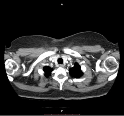
Figure 2: The mass in the right proximal metaphyseal humerus has the characteristic as a medullary lesion which had calcifications in computed tomography sections.

Figure 3: In coronal magnetic resonance images, the mass had low signal intensity on T1-weighted images

Figure 4: In axial magnetic resonance images, the mass had low signal intensity on T1-weighted images
X-ray was performed. There were multiple calcifications in proximal humerus on X-ray that was not found enough for the diagnosis (Figure 1). Therefore, computed tomography (CT), magnetic resonance imaging (MRI) and scintigraphy were performed. The mass in the proximal metaphyseal humerus has the characteristic as a medullary lesion which had calcifications in CT sections (Figure 2). In coronal (Figure 3) and axial (Figure 4) MR images, the mass had low signal intensity on T1-weighted images and heterogeneous high signal intensity on T2-weighted images. It had irregular nodular contrast enhancement and the mass did not cause the expansion of the bone. Increased activity was observed on scintigraphy.
Based on these findings, BS was assumed. The patient underwent follow up. In second month of follow up the growth in mass was seen on control MR images. So, an incisional biopsy was planned for the mass, because of the possible mimic of bone neoplasms. The biopsy was performed in operation room by minimal incision with the aid of C-arm fluoroscopy and the specimen was delivered to the pathology department. Pathological examination was reported as enchondroma.
Bone sarcoıdosıs (bs)
I Pathogenesis
Bone sarcoidosis, which is frequently a symptom of this multisystem disease, can also be associated with chronic and progressive disease biology. It was shown that those with bone involvement experienced a higher prevalence of three or more organ involvement compared to the sarcoidosis control group. Also, patients with BS were more likely to develop liver, spleen, or extra-thoracic lymph node involvement as well [18]. The pathogenesis of BS remains obscure. One theory hypothesizes that antigenic particles spread via blood and lymphatic vessels and compose granulomatous inflammations within the bone, bone marrow, and other organs. If this hematogenous scattering hypothesis is correct, this mechanism could explain the increased frequency of hematopoietic organ involvement like liver, spleen, or lymph nodes in the BS [18].
II Clinical findings
The prevalence of BS and specific bone involvement indicate a racial predisposition. In a study, it was observed that white patients were three times more likely than blacks to have BS, but black patients experience more hand bone involvement [18]. Small bones of the hands and feet are the more frequent sites of bone involvement with spine, pelvis, skull, and rib localizations rarely reported. On the other hand, axial bone involvement may be more than previously reported, because earlier studies relied mostly on plain X-rays, which may be less sensitive for axial bone lesions. In a study, it was observed that the most commonly affected site is axial skeleton, namely the spine (87.5%), followed by pelvis in BS. Hand involvement (15.6%) was the third most affected site, while no patient demonstrated foot involvement [18]. Similar to these findings, in another study, spine or pelvis involvement was reported in 90% of cases, while only 10% of cases revealed appendicular bone disease [19]. Probably, the more frequent availability of advanced imaging modalities as PET/CT and MRI in recent years may one of the reasons for this distribution change [18].
III Treatment
There is no general consensus for the treatment of BS. As in other disease symptoms, asymptomatic patients do not require treatment. However, as bone involvement is often indicative of multisystem disease, many patients should be treated for other organ manifestations. Corticosteroids are the most commonly prescribed first-line drugs in BS, but therapy is often limited by adverse effects. Methotrexate, as a steroid-sparing agent, has been reported useful for a variety of organ symptoms. Due to the long natural history of BS, the addition of methotrexate could act as a steroid-sparing agent to reduce toxicity. Hydroxychloroquine is also used in treating sarcoid patients, particularly for long term treatment. Azathioprine (second-line agent) and anti-TNF inhibitors (third-line treatment) are often prescribed for patients with chronic or severe sarcoidosis whose disease cannot be controlled by low dose corticosteroids [6, 18].
Discussion
BS is typically asymptomatic and is generally discovered incidentally. Sometimes it may cause pain that is a prominent feature of axial skeletal sarcoidosis, and it can be the initial site of disease. Also, reduced exercise capacity, numbness, swelling, or distortion of fingers may be symptoms of BS. Swelling and hand distortion are common features of appendiceal bone involvement [18]. Our patient was admitted to our clinic with right shoulder pain. But her shoulder's range of motion was within full limits. Bone marrow involvement in sarcoidosis may cause leukopenia and AST level may be elevated in liver involvement of BS [18]. In our case, her values of laboratory tests were all within normal limits except erythrocyte sedimentation rate (40mm/h) and C-reactive protein levels (50mg/L). Bone sarcoidosis typically causes nonspecific osteolytic lesions that can mimic neoplasms (e.g., osteoblastoma, metastases, or multiple myeloma), osteomyelitis, or bone cysts [5, 16]. In sarcoidosis involving the bone shaft like humerus and tibia, fractures may be more frequent due to pathologic disturbances and weakness in the bone architecture and there is concern that surgical repair will be difficult [5, 20, 21]. In this case, x-ray was performed first as radiological examination and were seen multiple calcifications in proximal humerus on X-ray. In sarcoidosis, bone lesions cannot usually appear on X-ray radiographs. In some cases, lytic lesions in the humerus and faint sclerotic lesion in the inferomedial humeral head were seen [22, 15]. Therefore, more sensitive and precise imaging modalities such as MRI are needed for the diagnosis [5].
Therefore, computed tomography (CT), magnetic resonance imaging (MRI) and scintigraphy were performed. The mass in the proximal metaphyseal humerus has the characteristic as a medullary lesion which had calcifications in CT sections. In BS, CT scanning has low sensitivity to small bones and appears insensitive for detecting sarcoid lesions in the hands and feet. In a study, positive CT imaging was seen in only 71.4% of 14 bone patients detected by other modalities. Although most patients in this bone affected group have evidence of intrathoracic sarcoidosis on chest imaging, normal chest X-rays and CT scans have been obtained from patients with documented spine involvement [18]. For that all, widespread vertebral, rib, and pelvic lytic lesions and focal area of mild sclerosis in the inferomedial humeral head can be seen in large bones [15].
We find that the mass had low signal intensity on T1-weighted images and heterogeneous high signal intensity on T2-weighted images in MR images. It had irregular nodular contrast enhancement and the mass did not cause the expansion of the bone. MRI studies are demonstrated multiple lytic bone lesions of humerus that simulated metastases or multiple myeloma; irregular signal in the upper humeral metaphysis including high signal intensity on T2 fat suppressed sequence and intermediate intensity on T1-weighted imaging with postcontrast enhancement in humeral metaphysis and abnormal marrow signal intensity in the medial humeral head, low signal intensity on both T1- and T2-weighted images, and a hyperintense signal focus within the marrow without cortical involvement in short tau inversion recovery (STIR) images [5, 15, 17]. Besides, involvement of cortical bone is less likely in these long bone lesions. Lack of cortical involvement may explain why many sarcoid lesions remain undiagnosed with radiographs in most cases. Sarcoidosis-related bone lesions resembling bone metastases on MRI may be the initial presentation. The presence of intralesional fat has been described as a feature that excludes malignancy [5, 7, 15]. Numerous areas of increased activity are seen positron emission tomography (PET) [17]. BS should be considered in sarcoidosis patients with bone lesions detected by either PET/CT or MRI. In these patients, biopsy may not be required because of the typical imaging patterns. In the presence of (-) chest X-ray findings, MRI or PET/CT scanning should be considered for the evaluation of sarcoidosis patients with unexplained bone complaints [18].
We observed increased activity on scintigraphy. In the literatüre, mild uptake in the humeral head can be seen 99mTc radionuclide scan [15]. Based on these findings, we assumed the BS and followed up her two month. In second month, the growth in mass was seen on control MR images. So, an incisional biopsy was planned for the mass, because of the possible mimic of bone neoplasms. Pathological examination was reported as enchondroma. Since the differential diagnosis of BS lesions includes also malignancy and infection, a biopsy may be required for confirmation. The presence of a non-caseating epithelioid granuloma in specimens is definitive evidence of BS [18].
Conclusıon
In here, we reported a rare case of enchondroma simulating BS of the proximal humerus. It was difficult to determine whether the patient's shoulder pain is actually caused by the lesion or another disease. Therefore, the sample was taken out by incisional biopsy from the lesion and confirmed to be enchondroma pathologically. Thus, the treatment of the patient's enchondroma was started on time. BS is a rare condition. We suspected the mass which was realized incidentally in a patient with sarcoidosis, could be BS. The biopsy revealed an enchondroma. Bone neoplasms should be kept in mind in cases like ours. Sarcoidosis can be a differential diagnostic challenge because of its nonspecific radiographic findings. Thus, a neoplastic cause such as primary osteoblastoma, metastasis, or multiple myeloma must always be excluded in differential diagnosis, together with other bone conditions such as osteomyelitis or bone cyst [7].
Conflicts of interest
Authors have no conflict of interest
Author’s contribution
The authors of this manuscript mentioned in the title contributed equally to the study and made substantial contributions to the following tasks of research: initial conception and design (K.B.); technical or material support (K.B.,A.A.); acquisition of data (S.S.); laboratory analysis and interpretation of data (N.S.); drafting of the manuscript (K.B.); critical revision of the manuscript for important intellectual content (K.B.,S.S.,N.S.) The views expressed herein are those of the authors and not necessarily their institutions or sources of support.
Article Info
Article Type
Case Report & Review of LiteraturePublication history
Received: Wed 22, May 2019Accepted: Fri 28, Jun 2019
Published: Tue 16, Jul 2019
Copyright
© 2023 Koray Basdelioglu. This is an open-access article distributed under the terms of the Creative Commons Attribution License, which permits unrestricted use, distribution, and reproduction in any medium, provided the original author and source are credited. Hosting by Science Repository.DOI: 10.31487/j.ACO.2019.02.03
Author Info
Corresponding Author
Koray BasdeliogluIstanbul Oncology Hospital Department of Orthopedics and Traumatology, Istanbul, Turkey
Figures & Tables
References
- Turcan A, Unel S, Sarıyıldız MA, Akkoyunlu ME (2012) Acute arthritis as initial presentation of sarcoidosis: Significance of chest X-ray. J Clin Experi Investiga 3: 102-104.
- Nishiguchi M, Furukawa F, Kanazawa N (2016) Leprosy versus sarcoidosis: Different diagnosis and review of misdiagnosed Cases. J Dermatolog Clin Res 4: 1087.
- Talmi D, Smith S, Mulligan EM (2008) Central skeletal sarcoidosis mimicking metastatic disease. Skeletal Radiol 37: 757-761. [Crossref]
- Soylu A, Türkmen M, Kasap B, Sarıoglu S, Saatci AO et al. (2004) Sarcoidosis with an uncommon presentation: apropos of a case. Turk J Pediatr 46: 366-369. [Crossref]
- Henrie TH, Skedros JG (2018) Sarcoid of the Upper Humerus Found Incidentally on MR Images Obtained for Work-Up of Rotator Cuff Tear Where Compromised Tissue Quality Was a Concern for Surgical Success. Case Rep Radiol 2018: 3579527. [Crossref]
- Nadera J. Sweiss, Elyse E. Lower, Peter Korsten, Timothy B. Niewold, Murry J. Favuset et al. (2012) Bone Health Issues in Sarcoidosis. Curr Rheumatol Rep 13: 265-272. [Crossref]
- Brandy-García AM, Cabezas-Rodriguez I, Caminal-Montero L, Suarez-Cuervo C, Redondo-Buil P (2017) Sarcoidosis mimicking lytic osseous metastases. Cleve Clin J Med 84: 753-754. [Crossref]
- Wilcox A, Bharadwaj P, Sharma OP (2000) Bone sarcoidosis. Curr Opin Rheumatol 12: 321-330. [Crossref]
- Chatham W (2010) Rheumatic manifestations of systemic disease: sarcoidosis. Curr Opin Rheumatol 22: 85-90. [Crossref]
- Ugwonali OF, Parisien M, Nickerson KG, Scully B, Ristic S et al. (2005) Osseous sarcoidosis of the hand: pathologic analysis and rewiev of the literature. J Hand Surg Am 30: 854-858. [Crossref]
- Waanders F, van Hengel P, Krikke A, Wesseling J, Nieboer P et al. (2006) Sarcoidosis mimicking metastatic disease: a case report and review of the literature. Neth J Med 64: 342-345. [Crossref]
- Cohen PR, Kurzrock R (2007) Sarcoidosis and malignancy. Clin Dermatol 25: 326-333. [Crossref]
- Barratt S, Burn PR, Stone R (2009) Osseous sarcoidosis masquerading as metastatic disease. Br J Hosp Med 70: 164-165. [Crossref]
- Costabel U, Oshimo S, Guzman J (2008) Diagnosis of sarcoidosis. Curr Opin Pulm Med 14: 455-461. [Crossref]
- Sidhartha Chaudhry, Michael L. Richardson (2006) Imaging Findings in Sarcoid of the Humerus. Radiol Case Rep 1: 154-158. [Crossref]
- Kuzyshyn H, Feinstein D, Kolasinski SL, Eid H (2015) Osseous sarcoidosis: a case series. Rheumatol Int 35: 925-933. [Crossref]
- Yachoui R, Parker BJ, Nguyen TT (2015) Bone and bone marrow involvement in sarcoidosis. Rheumatol Int 35: 1917-1924. [Crossref]
- Zhou Y, Lower EE, Li H, Farhey Y, Baughman RP et al. (2017) Clinical characteristics of patients with bone sarcoidosis. Semin Arthritis Rheum 47: 143-148. [Crossref]
- Sparks JA, McSparron JI, Shah N, Aliabadi P, Paulson V et al. (2014) Osseous sarcoidosis: clinical characteristics, treatment, and outcomes--experience from a large, academic hospital. Semin Arthritis Rheum 44: 371-379. [Crossref]
- Crawford B, Badlissi F, Lozano Calderón SA (2018) Orthopaedic Considerations in the Management of Skeletal Sarcoidosis. J Am Acad Orthop Surg 26: 197-203. [Crossref]
- Ungprasert P, Crowson CS, Matteson EL (2018) Risk of fragility fracture among patients with sarcoidosis: a population-based study 1976-2013. Osteoporosis Int 28: 1875-1879. [Crossref]
- Pallavi Mehrotra, Michelle Muller, Bernard Higgins, Ivan Zammit-Maempel (2011) An Unusual Cause of Widespread Lytic Bone Lesions Caused by Sarcoidosis. J Radiol Case Rep 5: 1-7. [Crossref]